Chapter 6 Annex 1: Species Profiles
In this section we provide additional reference material for top species covered in this report. Links to patent collections in the Lens database are provided to facilitate exploration of wider patent activity for the species.
6.1 Clarias macrocephalus
- Species name: Clarias macrocephalus
- Kingdom: Animalia
- Phylum: Chordata GBIF record
Brief Description of the Species:
Clarias macrocephalus lives in lowland wetlands and rivers - occurs in shallow, open water including sluggish flowing canals and flooded fields of the Mekong, and is capable of lying buried in mud for lengthy period if ponds and lakes evaporate during dry seasons. Clarias macrocephalus can move out of the water using its extended fins.37 It spawns in small streams between May and October.38
There are 5 catfish species found in Thailand with two of them being eaten by people: C. macrocephalus and C. batrachus. C. macrocephalus is reportedly considered to be the tastiest of the 2 by Thai people but is the more difficult of the two to cultivate.39 A hybrid created using C. macrocephalus females and males of the African C. gariepinus is farmed in Thailand on a large-scale and comes first in terms of farmed freshwater fish with a production of 86,475 tonnes or 30 percent of total production. It has recently been suggested production of the hybrid catfish is decreasing which may be due to the quality of the male African catfish which was introduced into Thailand a long time ago.40
C. macrocephalus is listed as near threatened by the IUCN - populations have been impacted by the loss of suitable wetland habitat through drainage and clearance for urbanisation and agriculture, as well as exploitation for aquaculture. In addition escaped hybrids from cultivation, are causing some concern.41
Known Distribution of the Species
Clarias macrocephalus is native to the Indochina peninsular: Vietnam, Cambodia and Laos as well as Thailand, and Malaysia.42.43 Clarias macrocephalus has also been recorded in Myanmar, Japan, China, Indonesia (Sumatra), Guam and the Philippines, but these are considered misidentifications or introductions .44 It is not commonly cultivated - although a hybrid of it is - but interest in farming it is on the increase.45

Figure 6.1: Photo of C.macrocephalus by FiMSeA, on http://ffish.asia
First description
Clarias macrocephalus, commonly known as the bighead catfish, was first described by Günther in 1864.46
6.1.1 Scientific Research
Research categories
Much of the research on Clarias macrocephalus in the region is categorised as Fisheries (20), Marine and Freshwater Biology (15), and Biochemistry & Molecular Biology (4).
Research summary
Clarias macrocephalus is an economically important fish in Thailand and is increasingly being cultivated in aquaculture settings (Na-Nakorn, Kamonrat, and Ngamsiri 2004). However C. macrocephalus doesn’t cultivate well and also has a near threatened status across its wild population range because of habitat loss; as such, much of the research effort is attempting to identify ways to increase production in aquaculture and minimise harm to the wild population.
A genetic population study examined the success of crosses between males of various strains of the fast growing African catfish C. gariepinus and females of strains of C. macrocephalus - which has tastier flesh - and found some hybrid strains had greater absolute growth rate than others (Koolboon et al. 2014). The use of hybrids has led to some conservation concerns in Thailand regarding the wild population of C. macrocephalus becoming back crossed with artificially created hybrids that have escaped from cultured systems. A study in Thailand took DNA samples from 25 natural populations of C. macrocephalus from across the country - they found 12 of the populations contained alleles in them from the African C. gariepinus (Na-Nakorn, Kamonrat, and Ngamsiri 2004). The hybrid is able to breed with both species which could lead to species extinction and also grows faster than C. macrocephalus and so might out-compete the native stock for resources (Na-Nakorn, Kamonrat, and Ngamsiri 2004).
Na Nakorn, et al., 1998, examined genetic distance in 4 wild populations in Thailand, they found two population groups, which were Chiangrai-Prachinburi and Pattani-Yala group and genetic distance was larger within the first group than those within the second group (Na-Nakorn et al. 1998).
Much of the research literature is focused on maximising yield in aquaculture settings through studying immunity to pathogens, as well as factors affecting growth and reproduction. Bacterial infection by Aeromonas hydrophila increased expression of the gene ‘HSC70-2’ in the tissues of C. macrocephalus which may relate to the role of HSC70-2 in the immune response of the species (Poompoung et al. 2012). Poompoung et al. (2012) found the C3 protein produced in the liver of C. macrocephalus had an essential role in immunity – furthermore an increase in C3 was induced by beta glucan in the diet of the fish.
The effect of stocking densities in aquacultures was investigated and found to be density dependent - C. macrocephalus fry reared at 285 m(-3) in tanks and at 114 m(-3) in ponds had significantly faster growth rates than fish reared at higher densities (Bombeo, Fermin, and Tan-Fermin 2002). Myostatin levels were found to have a negative feedback effect, rising levels reducing growth rate in larvae of C. macrocephalus when they were fasted for a period of time (Kanjanaworakul et al. 2014).
Methods of increasing reproduction, whilst minimising numbers of broodstock were investigated in the literature - as numbers of C. macrocephalus are limited. One study examined 5 forms of Gonadotropin releasing hormones to see the efficacy at stimulating ovulation in C. macrocephalus. Chicken GnRH was most effective as identical to catfish Gn RH (Ngamvongchon, Rivier, and Sherwood 1992). Another study looked at the success of using irradiated sperm of another fish species for gynogenesis - creating offspring with only the mothers DNA (Na-Nakorn, Kamonrat, and Ngamsiri 2004). Another study examined the properties required to create artificial seminal plasma to be able to dilute C. macrocephalus milt whilst still giving high fertilisation success of eggs -so less males are sacrificed during artificial insemination (Tan-Fermin et al. 1999). Clarias macrocephalus, harvested from irrigated rice fields in Malaysia, showed that spawning changes with water level (Ali 1993).
What to feed Clarias macrocephalus in culture was investigated by Bolivar and Fermin 1996, who found that catfish larvae can be given a dry diet, but growth rate is higher if live feed - artemia - is given prior to the dry formula diet (Fermin, Bolivar, and Gaitan 1996).
Aquatic or marine?
In a sector review of aquaculture in Thailand by the FAO Clarias macrocephalus and its hybrid with C. gariepinus are listed as freshwater species, not listed within the commercial brackish water production section of the report. There has been some discussion of the possibility of introducing some catfish species into brackish water aquaculture to make use of available coastal locations, but we didn’t find any reference to this having occurred for this species.47
Research locations
A conservation biology study in Thailand by Na-Nakorn, Kamonrat, and Ngamsiri (2004) took DNA samples from 25 natural populations of C. macrocephalus from across the country including- 12 populations from provinces located in the Chaophraya river basin in the centre of the country, 5 from the Mekong river basin, 1 from the east and 7 from the south of Thailand - they found 12 of the populations contained alleles in them from the African C. gariepinus (Na-Nakorn, Kamonrat, and Ngamsiri 2004). Another genetic study in Thailand OF C. macrocephalus, by Na Nakorn. et al in 1998, assayed 14 proteins across 4 wild populations from the north (Chiangrai), central (Prachinburi) and south (Pattani and Yala) of Thailand (Na-Nakorn et al. 1998).
In 1987, C. macrocephalus were sampled monthly from the rice fields, sump ponds and irrigation canals of North Kerian area up to about the latitude 5°45’, North western Peninsular Malaysia the fish are considered as one population (Ali 1993).
6.1.2 Patent activity
There are only 2 patent documents for C. macrocephalus. The most commonly cited patent document being for a specialized feed for sensitive organisms, especially larvae or juvenile forms of farmed aquatic organisms and the method of producing the feed WO2008084074A2.
The other C. macrocephalus patent document is for the use of vitamin K3 for treatment of parasitic disease in an individual, such as an animal or human being. Vitamin K3 has been found to be effective in the treatment of fish suffering from parasite infestation WO2009063044A1.
Search the titles, abstracts and claims of patent documents on the Lens or view the Lens public collection.
6.2 Lates calcarifer
- Species name: Lates calcarifer
- Kingdom: Animalia
- Phylum: Chordata GBIF record
Brief Description of the Species:
The Barramundi or Asian seabass (Lates calcarifer) is a widely distributed species of salt and freshwater sport fish, with large, silver scales, which can change shade, depending on their environments. L. calcarifer bodies can reach up to 1.8 m (6 ft) long, and the maximum weight is about 60 kg (130 lb). The average length is about 0.6-1.2 m (2–4 ft). It is very popular in Thai cuisine.48
Lates calcarifer are demersal, inhabiting coastal waters, estuaries, lagoons, and rivers; they are found in clear to turbid water, usually within a temperature range of 26−30 °C. This species does not undertake extensive migrations within or between river systems, which has presumably influenced establishment of genetically distinct stocks in Northern Australia.
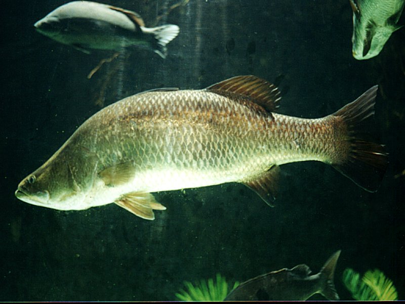
Figure 6.2: Lates calcarifer, Nick Thorne 2001, Creative Commons Attribution
Known Distribution of the Species
Lates calcarifer are a widely distributed species - from the Persian Gulf, through Southeast Asia to Papua New Guinea and Northern Australia.49
First description
Lates calcarifer was first described by German scientist Bloch, in 1790, and originally named Holocentrus calcarifer.50
6.2.1 Scientific Research
Research categories
Much of the research on Lates calcarifer is categorised as Fisheries (53), Marine and Freshwater Biology (45), Veterinary Sciences (13), Biotechnology & Applied Microbiology (9), Genetics & Heredity (8).
Research summary
Much of the research effort regarding L. calcarifer is concerned with optimising conditions for successful aquaculture of this valuable fish species. Some studies have investigated the natural history of the fish, particularly spawning and juveniles, as was the case in a study using controlled incubation of wild stock from the inland waters of Vietnam (Shadrin and Pavlov 2015).
Others have focused on optimising feed for this species – noting the success of live food in a study in the South China Sea (Shansudin et al. 1997). Others noted physiological aspects of nutrition – essential fatty acid metabolism in L. calcarifer (Mohd-Yusof et al. 2010). Some studies have trialled the use of local seaweed species - K. alvarezii, E. denticulatum and S. polycystum – to replace commercial feed binder in the feed of L. calcarifer juveniles (Shapawi and Zamry 2015).
The genetics of Lates calcarifer have been the subject of some investigation by scientists with one study looking at the chromosomes of male and female fish (Phimphan et al. 2015). Prevalent diseases which effect successful aquaculture were also given attention in the literature with one study noting the pathological effects of SDS in L. calcarifer indicating it to be a viral infection (Gibson-Kueh et al. 2011).
Fish aquaculture has an effect on the environment; this was the subject of a study examining pelagic carbon flow and water chemistry in mangrove estuaries with fish cage aquaculture of L. calcarifer (Alongi et al. 2003).
Aquatic or marine? Lates calcarifer live close to the sea floor, inhabiting coastal waters, estuaries, lagoons, and rivers; they are found in clear to turbid water - within a temperature range of 26−30 °C.51
This species is able to adapt to a range of salinities and thus are found in freshwater, estuarine, lagoons, brackish, rivers and coastal areas (Davis 1986). It has been shown that some of the fish move between salt and freshwater environments whilst others remain only in the marine environment (Davis 1986).
Adults in freshwater usually migrate to spawn in water with higher salinity as eggs need salt water to develop. Spawning most often occurs in brackish water like the river mouth and is seasonal (Moore 1982).
Research locations
Marine fisheries in the Gulf of Thailand were the subject of a study looking at changes in 35 marine species including Lates calcarifer over a 26 year period (Koolkalya, Sawusdee, and Tuantong 2015).
Mangrove inlets and creeks in Selangor, Malaysia are the habitat for 119 species of fish (the majority of which are juveniles)- the common fish species included Lates calcarifer (Sasekumar et al. 1992). The tidally dominated mangrove estuaries of peninsular Malaysia were the location of a study examining the environmental effects of fish cage aquaculture of L. calcarifer (Alongi et al. 2003).
6.2.2 Patent activity
The majority of patent activity centres around aquaculture, specifically, feed (WO2008084074A2), products (WO2010027788A1) and methods(WO2009063044A1).
Patent activity includes the novel use of fish skin including Lates calcarifer as an industrial source of collagen (see US20120114570A1, US20120114570A1).
Medical and cosmetic skin treatments have also been derived from the hatching fluid of fish including Lates calcarifer (WO2014094918A1).
Search the titles, abstracts and claims of patent documents for this species on the Lens or view the Lens public collection.
6.3 Litopenaeus vannamei
- Species name: Litopenaeus vannamei
- Kingdom: Animalia
- Phylum: Arthropoda GBIF record
Brief Description of the Species:
The dominant shrimp culture species in the world, they are benthic - occurring in waters of depth between 0-72m in marine habitats as adults, and estuarine environments as juveniles in subtropical to tropical climates.52
Asia has seen a phenomenal increase in the production of L. vannamei, no production was reported to FAO in 1999, but by 2004 it was nearly 1,116,000 tonnes having overtaken the production of P. monodon in China, Taiwan Province of China and Thailand. However, due to fears of importing exotic diseases it is only experimentally farmed in Cambodia, India, Malaysia, Myanmar and the Philippines. Thailand and Indonesia both freely permit its commercial culture but have official restrictions, so that only SPF/SPR broodstock may be imported.53
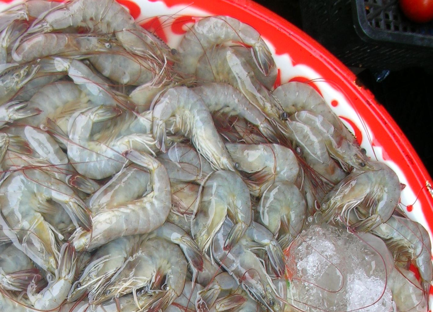
Figure 6.3: Litopenaeus vannamei, By Xufanc (Own work)
Known Distribution of the Species
Litopenaeus vannamei is native to the Eastern Pacific coast from Sonora, Mexico in the North, through Central and South America as far South as Tumbes in Peru.54
The main producer countries of Litopenaeus vannamei are: China, Thailand, Indonesia, Brazil, Ecuador, Mexico, Venezuela, Honduras, Guatemala, Nicaragua, Belize, Vietnam, Malaysia, Taiwan P.C., Pacific Islands, Peru, Colombia, Costa Rica, Panama, El Salvador, the United States of America, India, Philippines, Cambodia, Suriname, Saint Kitts, Jamaica, Cuba, Dominican Republic, Bahamas.55
First description
Litopenaeus vannamei was first described in 1931 as Penaeus vannamei by Boone.56
6.3.1 Scientific Research
Research categories
Much of the research on Litopenaeus vannamei is categorised as Fisheries (54), Marine and Freshwater Biology (32), Veterinary Sciences (26), Biotechnology & Applied Microbiology (10), Immunology (10), Biochemistry and Molecular Biology (6).
Research summary
Much of the research on Litopenaeus vannamei as the worlds dominant cultured shrimp species was with regard to maximising yield, through selecting robust broodstock as well as via disease treatment and prevention.
Researchers found that cold storage of spermatophores from broodstock was effective by combining an antibiotic within the mineral oil surrounding the sperm (Koolkalya, Sawusdee, and Tuantong 2015). Another study focussed on reproduction of Litopenaeus vannamei looked at the success of crosses with limited numbers of males and females, and noted that male broodstock could be selected with desirable traits (Aungsuchawan et al. 2008).
Diseases of Litopenaeus vannamei and other commercially farmed shrimps blight production and so much of the research literature is focussed on how to overcome some of these diseases: via longer rearing and less dense stocking to protect against infectious myonecrosis Silva et al. (2010), or introduction of immune stimulating agents PAP-phMGFP and CHH (Silva et al. 2010; Khimmakthong et al. 2011; Wanlem et al. 2011). Another study found the use of dsRNA injected into prawns to be effective as both a preventative, and treatment for Penaeus stylirostris densovirus (PstDNV) infection (Ho et al. 2011). Similarly galangal extract and trans-p-coumaryl diacetate stimulated the immune system response, thereby promoting resistance to V. harveyi infection in Litopenaeus vannamei (Chaweepack et al. 2014). Detecting Taura Syndrome Virus in Shrimps via diagnostic test kits was the subject of another study (Chaivisuthangkura et al. 2006).
Antibiotic resistance is a growing problem worldwide in the aquaculture industry and for human health. As such much of the literature was focussed on looking at alternatives to antibiotic use, such as probiotics. One study found the use of bacteria, yeasts, and algae in water additives or artemia enriched enhanced survival and growth rate of Litopenaeus vannamei (Nimrat, Boonthai, and Vuthiphandchai 2011). The probiotic Bacillus subtilis BS11 supplement was found to benefit Litopenaeus vannamei growth and survival after infection with Vibrio harveyi, conferring disease resistance (Sapcharoen and Rengpipat 2013).
The food safety of cultured seafood is paid some attention amongst the research literature, -minimal sequential ozone treatment and ice storage for Litopenaeus vannamei has been shown to have an antioxidant like action and could promise further safety for shrimp consumption (Okpala 2014).
Astaxanthin extract from Litopenaeus vannamei has been demonstrated in one study to act as anti-inflammatory alternative against carrageenan-induced paw oedema and pain behaviour in mice (Kuedo et al. 2016).
Genetics were the subject of several studies in the literature including the correlation between genetics and body weight in Litopenaeus vannamei (Glenn et al. 2005). Genetic variation and population structure of wild Litopenaeus vannamei from 4 geographic locations from Mexico to Panama were investigated using 5 microsatellite DNA and it was found that sub-populations exist (Valles-Jimenez, Cruz, and Perez-Enriquez 2004).
Aquatic or marine? Litopenaeus vannamei live in marine habitats as adults, and estuarine environments (brackish water) as juveniles in subtropical to tropical climates.57
Research locations
Brazil and Indonesia are mentioned in a study on infectious myonecrosis of Brazilian farmed Litopenaeus vannamei (Silva et al. 2010).
China, Thailand, Peru to Mexico on the Pacific coast, and Taiwan are referenced in a paper about the most widely cultured alien crustacean in the world Litopenaeus vannamei (Liao and Chien 2011). Genetic variation among 4 populations of L. vannamei from locations along the coast between Mexico and Panama were the subject of one investigation which noted sub populations were present (Valles-Jimenez, Cruz, and Perez-Enriquez 2004).
Enterocytozoon hepatopenaei has been found in L. vannamei farms across India - within the states of: Tamil nadu, Andhra Pradesh and Odisha. E. hepatopenaei has also been detected in shrimp cultured in China, Vietnam and Thailand and is suspected to have occurred in Malaysia and Indonesia and to be linked with severely retarded growth (Biju et al. 2016).
6.3.2 Patent activity
There are 309 patent documents for Litopenaeus vannamei, the most commonly cited patent document is for a commercial aquaculture system using algae and artemia as food stuffs for juveniles US6615767B1. Many other patent documents relate to differing forms of aquaculture system for growing Litopenaeus vannamei. Other L. vannamei patent documents relate to feed compositions, such as one without fish meal CN102648738A.
Some patent documents for L. vannamei relate to inducing immune responses, for example using dsRNA to modulate gene expression and responses to pathogens US20050080032A1.
Search the titles, abstracts and claims of patent documents for this species on the Lens or view the Lens public collection.
6.4 Macrobrachium rosenbergii
- Species name: Macrobrachium rosenbergii
- Kingdom: Animalia
- Phylum: Arthropoda GBIF record
Brief Description of the Species:
Macrobrachium rosenbergii known as the Giant River Prawn and the Giant Freshwater Prawn - it is widely fished and also is the main freshwater shrimp cultivated within the commercial aquaculture industry.58 The body length of this large prawn species is 320 mm for males; and 250 mm for females, as it is a sexually dimporphic species.59
Global production of the Giant River Prawn has grown since its inception in the 1970’s and by 2002 was 200 000 tonnes/yr.60
Known Distribution of the Species
M. rosenbergii is native to Bangladesh; Brunei Darussalam; Cambodia; China; India; Indonesia (Java); Malaysia (Peninsular Malaysia, Sabah, Sarawak); Myanmar (Myanmar (mainland)); Pakistan; Philippines; Singapore; Sri Lanka; Thailand.61 The geographic distribution is now much wider than this as it has been introduced to all continents for aquaculture and is now found in Brazil, Venezuela, Australia, United States, Iceland, Japan, Germany, Iran, Taiwan.62
First description
M. rosenbergii was first described by Johannes De Man in 1879 as Palaemon rosenbergii in the publication: De Man, 1879, Notes Leyden Mus., 1:167, however in 1966 Johnson renamed it to its current nomenclature of Macrobrachium rosenbergii.63
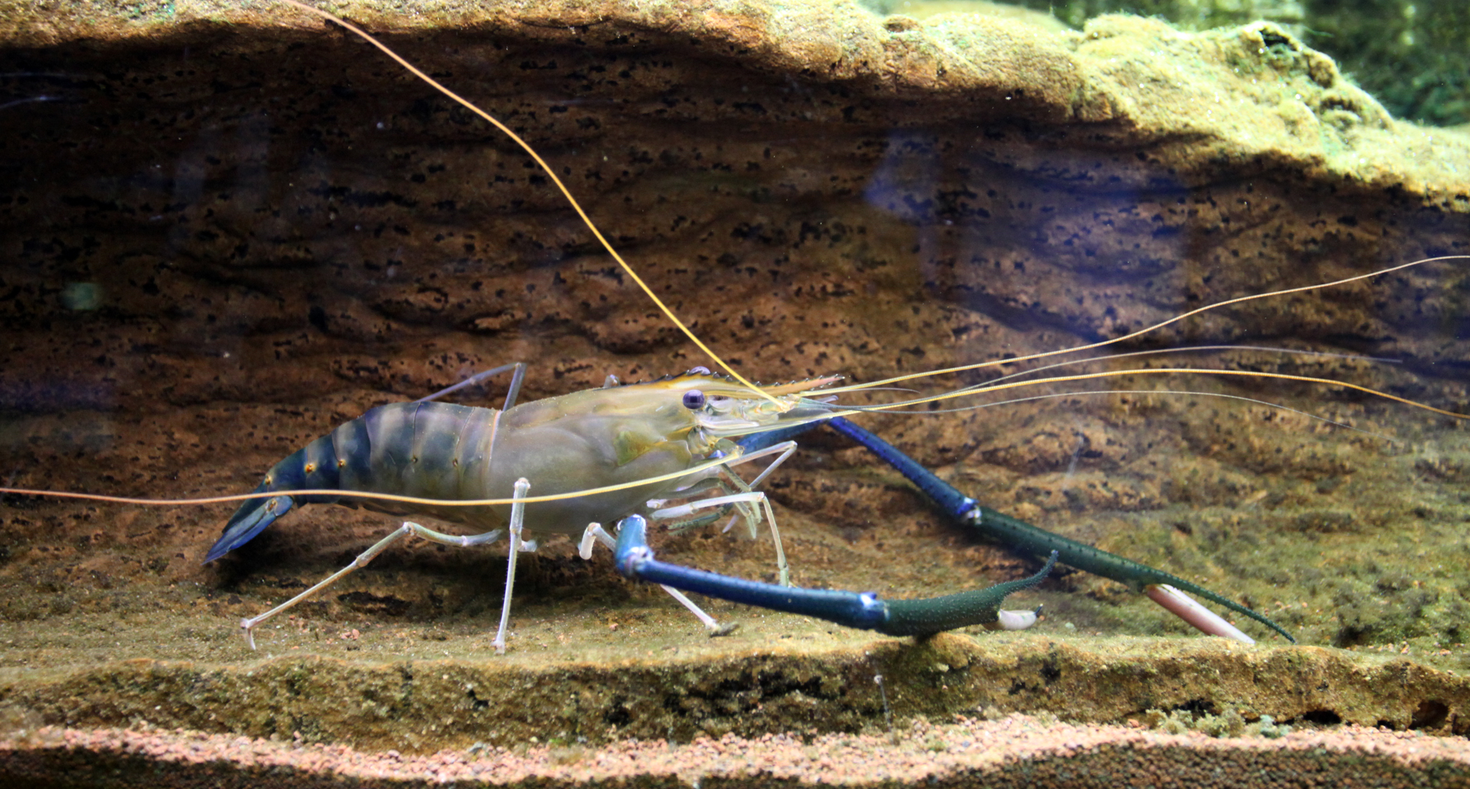
Figure 6.4: Macrobrachium rosenbergii, By Citron CC BY-SA 3.0
6.4.1 Scientific Research
Research categories
Much of the research on M. Rosenbergii in the region is categorised as fisheries (77), followed by Marine and Freshwater Biology (46), Biochemistry & Molecular Biology (21), Veterinary Sciences (20), Zoology (13), Immunology (12) amongst others.
Research summary
The majority of the research effort on Macrobrachium rosenbergii is directed towards maximising aquaculture yield to increase economic gain of shrimp farmers. So the research is broadly concerned with disease prevention/treatment, nutrition, stocking densities, reproductive success, and genetic make up of broodstock. Understanding the diseases of M. rosenbergii - such as L. garvieae a bacteria that infects shrimps (Dangwetngam, M. and Suanyuk, N. 2014), and White Tail Disease virus which causes large scale mortality in aquaculture populations - can enable treatments and preventative strategies to be adopted (Bonami and Widada 2011). Improving yield of shrimp farmers has led many to look to the genetic quality of broodstock - analysing the parentage of existing broodstock, as well as comparing this with wild samples (Karaket and Poompuang 2012; Thanh et al. 2015). This genetic analysis can then help develop a more successful breeding strategy (Thanh et al. 2010).
The nutritional composition of feed given to farmed M. rosenbergii is another common theme of research in the literature, as well fed shrimps grow bigger and reproduction is increased (Kangpanich et al. 2016). However for shrimp farmers in the Mekong Delta region of Vietnam, identifying non commercial feed options - due to affordability issues- is key (Hien et al. 2005). Understanding how eye stalks inhibit reproduction in female M. rosenbergii, as well as understanding how sex differentiation occurs and when to intervene to create all male stock are some of the reproductive focussed areas of research covered in the literature (Sripiromrak et al. 2014; Jung et al. 2016; Rungsin, Swatdipong, and Na-Nakorn 2012).
Aquatic or marine? Although M. rosenbergii is predominantly a tropical freshwater species, the larval stage of the species is found within adjacent brackish water, as females migrate to estuaries to lay their eggs.64 Most of the M. rosenbergii farms are in freshwater inland locations, however some are in estuarine and coastal locations where salinity in the ponds can fluctuate and effect breeding success of females (Yen and Bart 2008).
Research Locations
Much of the research involves laboratory bred cultures of M. rosenbergii, although some studies do include wild samples as is the case in a genetic study by Schneider et al. (2012, 2012). Schneiders’ study used cultured prawn samples from: Andhra Pradesh, India; Kaneohe, Hawaii, USA; Tamashiro Market, Honolulu, Hawaii, USA; University of Negev, Beer Sheva, Israel; Frankfort, Kentucky, USA; Leland, Mississippi, USA; Weatherford, Texas, USA as well as wild prawn samples from Hmaw River, Hlaing River and the Pan Hlaing River - tributaries of the Yangon River, Yangon, Myanmar; and Mahanadi River, Orissa, India. There are some references to more specific locations for wild samples within the literature - such as research into a breeding strategy for genetic improvement using wild strains Vietnam (Dong Nai and Mekong) (Thanh et al. 2010; Tidwell et al. 2014). L. garvieae was isolated from M. rosenbergii samples taken from cultures and wild prawns from the Phatthalung and Songkhla, provinces of southern Thailand (Dangwetngam, M. and Suanyuk, N., 2014).
Some research doesn’t specify anything more than the country such as in a study detailing culture technology in Thailand (Na-Nakorn and Jintasataporn 2012). Genetic diversity analysis was undertaken on cultured samples from farms in Zhejiang, Guangdong and Guangxi provinces, China, as well as wild samples from Dong Nai River and Mekong River, Vietnam (Thanh et al. 2015).
6.4.2 Patent activity
The top cited patent involving M. rosenbergii is regarding feed composition for shrimps in aquaculture settings WO2008084074A2. The invention specifies a range of percentage compositions of lipid:carbohydrate:protein, differing nutritional sources, particle size and types - as an alternative to live feed for shrimp larvae.
The largest family for a patent application associated with M. rosenbergii is for another type of feed, this time for preparation and use of methionylmethionine as a feed additive for fish and crustaceans US20100098801A1.
Search the titles, abstracts and claims of patent documents for this species on the Lens or view the Lens public collection.
6.5 Nypa fruticans
- Species name: Nypa fruticans
- Kingdom: Plantae
- Phylum: Tracheophyta GBIF record
Brief Description of the Species:
Nypa fruticans is a palm found in the upstream estuarine zone in all intertidal regions. It forms extensive belts along brackish to tidal freshwater creeks and rivers. It is very fast growing, especially in fresh water, and is a competitive species.65 It is the only palm adapted to the mangrove biome and grows in terrestrial, marine and freshwater habitats.
The trunk of Nypa fruticans grows underground with the leaves, female flower inflorescences and male flowers visible above the ground.66
Nypa fruticans is used for a wide range of commercial goods and services including- thatching and for making alcoholic drinks through a fermentation process.67 It is an important emerging source of biofuel, capable of producing ethanol ranging from 6,480 to 20,000 L/ha, a higher yield than sugar cane.68 It is considered to be of ’Least concern as it is widespread and locally common.69
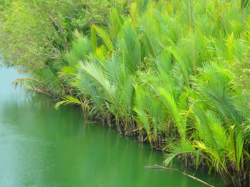
Figure 6.5: Nipa palms, By Qaalvin, own work
Known Distribution of the Species
Nypa fruticans ranges from Sri Lanka and the Ganges Delta through to the west Pacific. In South and South-East Asia it is found in Bangladesh, Brunei Darussalam, Cambodia, China, India, Indonesia, Japan, Malaysia, Myanmar, Singapore, Sri Lanka, Thailand, and Viet Nam . In much of its native range it has been planted and exists in large or small-scale plantations.70
Nypa fruticans has been introduced to Cameroon and Nigeria in West Africa and to Panama in Central America and Trinidad and Tobago in the Caribbean.71
First description
Nypa fruticans was first described by a German botanist called Christoph Carl Friedrich von Wurmb in 1781.72
6.5.1 Scientific Research
Research categories
Much of the research on Nypa fruticans is categorised as Environmental Sciences (8), Biology (6), Marine and Freshwater Biology (6), and Plant Sciences (6).
Research summary
Much of the research on Nypa fruticans is studying its role within mangrove biomes, an important ecological and economic habitat type. In the Segara Anakan lagoon, Java, mangrove tree density and diversity was examined at the eastern part of the lake - Nypa fruticans was present and characterised a mature undisturbed forest (Hinrichs, Nordhaus, and Geist 2008). Within Malaysia - on Carey Island - a similar demographic study was conducted looking at the percentage of Nypa fruticans seedling, juvenile, adult and mature present the population showed a majority of adults with 67.9% (Nasrin and Z 2010). Also in Malaysia a study of forest structure, diversity index and above-ground biomass was conducted on N. fruticans at Tok Bali mangrove forest, Kelantan (I, J, and I 2007).
Low genetic diversity for N. fruticans was found amongst six natural populations from China, Vietnam, and Thailand across a total of 183 individuals (Jian et al. 2010). Creating N. fruticans seedlings was the focus of some research due to the economic benefits of the plant - mass clonal propagation of disease free planting materials was successfully trialled in one study (JA et al. 2015). In another propagation study in Vietnam, a participatory action research methodology was used to successfully establish an N. fruticans (and other mangrove species) nursery on unused acid sulphate soil - normally considered unsuitable for mangrove growth (T. Nguyen et al. 2016).
Species living on N. fruticans or within the mangrove biome it creates were the focus some of the literature with several studies examining the fungi present on samples from different populations. One study tested the fungi growing on N. fruticans for heavy metal tolerance and found that one that was able to successfully grow deposit heavy metal pollution - meaning it could potentially be used as a type of absorbent material (Choo et al. 2015). Species diversity of marine fungi on Nypa fruticans in Samut Songkhram Province, Thailand were investigated, with 81 fungal taxa recorded (Pilantanapak, Jones, and Eaton 2005). The taxonomy of fungus found on Nypa fruticans in the intertidal regions in Trang and Trat provinces, Thailand was examined in another study (Suetrong et al. 2015). Biodiversity and ecology of higher filamentous fungi on Nypa fruticans along the Tutong River, Brunei were examined during 1999, with Forty-six taxa recorded (KD and VV 2006).
Both mudskippers and fireflies were found to inhabit populations of Nypa fruticans within the literature. Mudskippers, Periophthalmodon septemradiatus were recorded for the first time in Peninsular Malaysia, from the small tributaries of the rivers Selangor and Muar (MZ and Y 2003). Whereas Pteroptyx fireflies are commonly reported to congregate in large numbers in mangroves, but researchers found they preferred vegetation consisting mainly of S. caseolaris and N. fruticans (Jusoh, Hashim, and Ibrahim 2010). A study of the root-associating bacteria of N. fruticans found in brackish-water mud in Sarawak, Malaysia, discovered that Burkholderia vietnamiensis to be its main nitrogen-fixing bacterium (Tang et al. 2010). Another important type of organism examined in the literature was that of the pollinators inhabiting N. fruticans mangroves in an Oxford University study, within the submerged Melaleuca forests of Vietnam (Tan 2008).
The economic exploitation of N. fruticans was examined by some researchers, one Malaysian paper looked at the ripe and unripe fruit content to see if can be used as a food source (Sum, Khoo, and Azlan 2013). Whilst another scientist examined the potential of N. fruticans in the Philippines for alcohol production (Jr 2010).
Aquatic or marine?
N. fruticans grows in mangrove forests in upwater estuarine locations, so it is aquatic, and terrestrial - as it is exposed at low tide, and marine as the salt water from the ocean meets the freshwater of the rivers.73
Research locations
A demographic study of Nypa fruticans was undertaken on Carey Island Malaysia (Nasrin and Z 2010). Forest structure was also examined at Tok Bali mangrove forest, Kelantan, Malaysia Kamaruzaman (I, J, and I 2007). A study looking at tree density and diversity of mangroves in different areas of Segara Anakan lagoon, Java, noted the presence of N. fruticans as an indicator of a mature forest (Hinrichs, Nordhaus, and Geist 2008).
In the Vam Ray area Kien Giang Province, Vietnam, a participatory research methodology was used to establish a mangrove nursery on soil considered previously to be unsuitable - 5 types of mangrove species were successfully grown including Nypa fruticans seedlings; providing a potential source of income for local people (H. Nguyen et al. 2016).
Genetic diversity of populations from six natural populations of Nypa fruticans from China, Vietnam, and Thailand was assessed, and showed an extremely low level of genetic diversity (Jian et al. 2010).
Selangor and Muar rivers of Malaysia were discovered to contain mudskipper populations, whilst a 2010 study of alcohol production potential took place in Vinzons, Camarines Norte, Philippines (Jr 2010). Pollinators of Nypa fruticans were examined in the submerged meleuca forests of South Vietnam in one study and in another and the bacteria inhabiting the rhizomes of Nypa fruticans in Sarawak Malaysia were identified (Tan 2008; Tang et al. 2010).
6.5.2 Patent activity
There are 10 patent documents for Nypa fruticans, the most commonly cited document is for a tnf-alpha and nitric oxide production inhibitor using an extract from the plant body of the palm JP2007008817A.
Search the titles, abstracts and claims of patent documents for this species on the Lens or view the Lens public collection.
6.6 Oreochromis niloticus
- Species name: Oreochromis niloticus
- Kingdom: Animalia
- Phylum: Chordata GBIF record
Brief Description of the Species:
Oreochromis niloticus is a species of Tilapia, a cichlid fish native to the Nile basin in Africa and coastal rivers of Israel, however it’s an economically important species of fish which has been introduced widely outside of its natural range.74
O. niloticus adults grow to 60cm and live up to 9 years weighing up to 4.3kg. It lives in freshwater and can tolerate brackish water, with a temperature range of 8-42°C. It’s an omnivore feeding on plankton as well as mosquito larvae. Groups of cichlids establish social hierarchies in which dominant males have priority for access to mate and food.
Commercial aquaculture of O. niloticus dates back to ancient Egypt, as they are fast growing and produce good fillets. The wild type of O. niloticus is less popular today as the meat is dark so a variety with lighter meat is used in aquaculture.
Known Distribution of the Species
O. niloticus has been taken from its native African and Israeli habitats to tropical and subtropical waters all over the world for aquaculture. Nile tilapia from Japan were introduced to Thailand in 1965, and from Thailand they were sent to the Philippines. Nile tilapia from Cote d’Ivoire were introduced to Brazil in 1971, and from Brazil they were sent to the United States in 1974. In 1978, Nile tilapia was introduced to China, which leads the world in tilapia production and consistently produced more than half of the global production in every year from 1992 to 2003.75
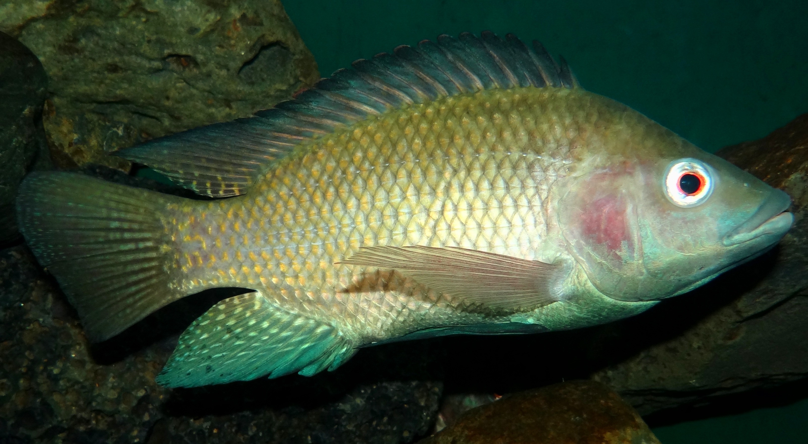
Figure 6.6: Oreochromis niloticus, By Bjørn Christian Tørrissen, own work
First description
Oreochromis niloticus was first described by Linnaeus in 1758 who named it Perca nilotica, this was changed to the current name by Greenwood, P. H. 1960 in his publication which revised the Lake Victoria Haplochromis species.76
6.6.1 Scientific Research
Research categories
Much of the research on Oreochromis niloticus is categorised as Fisheries (78), Marine and Freshwater Biology (61), Veterinary Sciences (14), Agriculture, Dairy and Animal Science (11).
Research summary
The research literature on Oreochromis niloticus is largely concerned with improving productivity of the aquaculture industry that grows tilapia commercially. Methods of aquaculture are investigated: such as factors effecting rice polyculture, brackish water aquaculture and cage aquaculture. Rice polyculture involving the farming of Oreochromis niloticus amongst other fish species in paddy fields has several elements for investigation including rice seeding rate, nitrogen cycling and the ecological/economic benefits of this form of polyculture (Vromant, Nam, and Ollevier 2002; Rothuis et al. 1999; Oehme et al. 2007).
Brackish water aquaculture requires salinity tolerance, which is not a feature of Oreochromis niloticus - it is the least salt tolerant tilapia, but has a quick growth rate. O. niloticus is increasingly being cultured in coastal ponds (especially in the Philippines, Indonesia and Vietnam) - sometimes with shrimps - so it would be beneficial if a more salt tolerant hybrid could be produced (Kamal and Mair 2005).
Stocking density of O. niloticus in cages was investigated by the Asian Institute of Technology in Thailand (Yi, Lin, and Diana 1996). The combined net yield of both caged and open-pond tilapia was found to be highest in the treatment with 50 fish m(-3).
The feed used in the aquaculture of O. niloticus is the focus of much research effort, including the replacement of fish oil with vegetable oil - which gave worse yields, however using cheap farm waste -mushroom stalks - to replace rice bran in feeds gave a greater yield for less money (Karapanagiotidis et al. 2007; Bahari et al. 2015). Scientists supplementing O. niloticus feed with antimicrobial herbal extracts found no mortality in S. agalactiae infected Nile tilapia for fish receiving dried matter of A. paniculata aqueous extract supplemented feeds at ratios (w/w) of 4:36 and 5:35 (Rattanachaikunsopon and Phumkhachorn 2009). Probiotic supplemented feeds were found to increase survival in O. niloticus infected with Aeromonas hydrophila (Kaew-on, Areechon, and Wanchaitanawong 2016).
Infections of fish with pathogens in intensively farmed aquaculture environments can lead to mass mortality and economic loss, hence much of the literature is concerned with understanding pathogens, and methods of combating losses. Intensive O. niloticus egg incubation creates conditions favourable for microbe growth leading to mass mortalities of fish larvae (Jantrakajorn and Wongtavatchai 2016). Scientists studied the immune response of O. niloticus to better understand how to develop vaccines (Natthaporn et al. 2015). Occurrence of infections in cultured populations of O. niloticus are studied to better understand the disease as was the case with a population infected with Neoechinorhynchus in the Philippines (Cruz CPP and VGV 2012).
Heavy metal contamination of watercourses containing farmed O. niloticus is a concern in the literature due to heavy industry waste being released, an and the potential resultant build up in the flesh of farmed fish eaten by local people (Baharom and Ishak 2015; Marcussen et al. 2007). Reduction of copper-induced tissue changes in O. niloticus by calcium exposure is beneficial in reducing effects in marine species (Kosai et al. 2009).
Many O. niloticus farmers produce all-male populations because of the superior growth rate of males compared to females. The literature discussed different methods for achieving this such as hormonal sex reversal, as well as genetic manipulation to create YY male broodstock achieving a 95.6% male sex ratio in the population (Guerrero 2008; Mair et al. 1997).
Understanding the genetic make up of different breeds of O. niloticus, and selective breeding using different strains to genetically improve broodstock - is the focus of some researchers (Supiwong et al. 2013; Bentsen et al. 1998). Red Tilapia, a strain of O. niloticus bred for its white flesh, is a popular strain for culture.
Aquatic or marine?
Native to the Nile basin rivers and lakes in Africa and coastal rivers of Israel] O. niloticus is primarily a freshwater fish.77 However brackish water aquaculture is on the rise in Philippines, Indonesia and Vietnam which culture O. niloticus - which requires salinity tolerance - not a strength of Oreochromis niloticus, as it is the least salt tolerant cichlid fish, but has a quick growth rate; research is being conducted to breed a more salt tolerant hybrid of Oreochromis niloticus (Kamal and Mair 2005).
Research locations
O. niloticus is farmed in its native African countries, and Israel but has also been exported for aquaculture in fresh and brackish water locations across the world including: Egypt, Thailand, Philippines, Indonesia, Malaysia, Vietnam, and Bangladesh.
Some more specific locations are mentioned in the literature such as a study sampling the fish pathogen Aeromonas hydrophila occurrence in the O. niloticus population in West Bay of Laguna de Bay, Barangay Bayanan, Muntinlupa City, Malaysia in three months during 2005 (R. Rodriguez et al. 2006). Similarly another study sampled a population in Sampaloc Lake, Philippines for the occurrence of a fish pathogen called ‘Neoechinorhynchus’ (Cruz CPP and VGV 2012). Polyculture of O. niloticus with ‘bleeker’ was the focus of a study in the Mekong delta Vietnam. Heavy metal incidence was investigated in two separate studies in Indonesia one sampling the Galas River & Beranang mining pool, Selangor and another sampling Lake Cirata, West Java (Baharom and Ishak 2015; Salami et al. 2008). Nitrogen cycling in rice fish culture in Bangladesh, was the subject of a study (Oehme et al. 2007). Spawning season was studied in two lakes in the Cote d’Ivoire (Duponchelle et al. 1999). The spatio-temporal dynamics of fish larvae in Sirindhron Reservoir, Lower Mekong Basin, north-east Thailand (Jutagate et al. 2016).
6.6.2 Patent activity
A partitioned aquaculture system for O. niloticus and other marine organisms, with an algal channel to control flow rate in the system US_6192833_B1 is the invention associated with O. niloticus that has been cited most often.
The preparation and use of methionylmethionine as feed additive for fish and crustaceans US_2015_0223495_A1 is the patent document associated with O. niloticus with the largest patent family.
Search the titles, abstracts and claims of patent documents for this species on the Lens or view the Lens public collection.
6.7 Penaeus mondon
- Species name: Penaeus monodon
- Kingdom: Animalia
- Phylum: Arthropoda GBIF record
Brief Description of the Species:
Penaeus monodon, the Giant Tiger Prawn, is one example of a group of giant prawn species with economical importance to the commercial fisheries industry.78 The total catch reported for this species for 1999 was 144 042 t with the largest catches from India (93 830t) and Indonesia (31 510 t).79
P. monodon lives in marine (adults) and estuarine (juveniles) environments, between 0-110 metres in depth and grow to a maximum of 336mm in length, weighing 60-130g.80
There are two specimens of P. monodon recorded on Vertnet, both are held at the University Museum of Zoology Cambridge and were collected in India - one specifies the Madras region.81
Known Distribution of the Species
“Indo-West Pacific: E. and S.E. Africa and Pakistan to Japan, the Malay Archipelago and northern Australia.”82
First description
Penaeus monodon was first described by Johan Christian Fabricus in 1798, but clarification of which species this name referred to came later in 1949, when Lipke Holthuis showed it to be the type species of the genus Penaeus.83
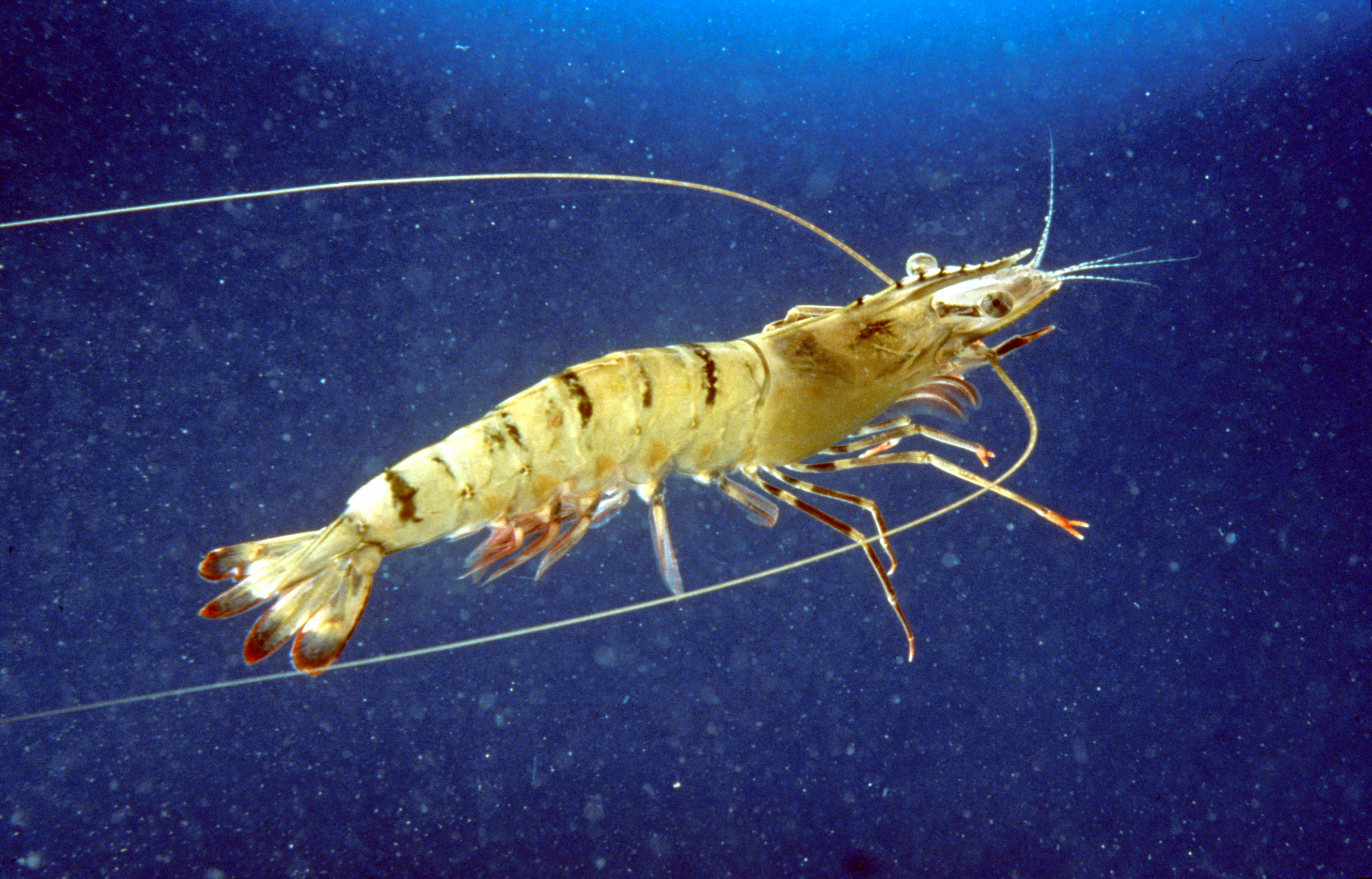
Figure 6.7: Penaeus monodon, CSIRO
6.7.1 Scientific Research
Research categories
Much of the research on P. monodon is categorised as fisheries (288), followed by Marine and Freshwater Biology (179), Veterinary Sciences (123), Immunology (81), Zoology (60), Biochemistry & Molecular Biology (58), Biotechnology and Applied Microbiology (45), Virology (32) and so on.
Research summary
The majority of the research effort on P. monodon is directed towards maximising fisheries yield and increasing economic gain. As such a lot of the P. monodon literature is concerned with understanding the immune response of the shrimp populations to the main viral and bacterial pathogens which cause large scale mortality in the population, such as White Spot Syndrome Virus (WSSV), Yellow Head Virus (YHV) and Vibrio harveyi (Ponprateep, Tassanakajon, and Rimphanitchayakit 2011; Jaree, Tassanakajon, and Somboonwiwat 2012). Whilst others are focussed on improving shrimp reproduction for example injecting female broodstock with a hormone that stimulates the ovaries (Sathapondecha, Panyim, and Udomkit 2015). Some work is concerned with identifying sub-populations of P. monodon where there is significant genetic differentiation between samples collected from different geographic areas (P et al. 2000).
Aquatic or marine?
P. monodon is fished from wild populations in offshore marine waters, however there is reference to inshore and pond fishing in some countries: Singapore, Malaya, Philippines, Thailand and Vietnam (Engle et al. 2017).84 In addition juveniles live in river mouth and estuarine mangrove environments predominantly - which act as a nursery with adults living in deeper offshore environments. Wild marine populations are commonly used to establish disease free broodstock for commercial pond fisheries inland (Claydon et al. 2010). Thus some of the research is solely focused on captive inland populations from commercial farms whilst others are comparative studies between wild and captive specimens (Caipang and Aguana 2010).
Research locations
Much of the research involves laboratory bred broodstock of P. monodon, but from different geographical populations, where often only the country is cited: Australia, China, Taiwan, Malaysia, Vietnam, Thailand, Papua New Guinea, New Caledonia, India, Singapore, Philippines, Tanzania, Japan, Sulawesi, Sumatra, and Brazil. There are some references to more specific locations within the literature such as research in the Mekong Delta Region of Southern Vietnam looking at different pond farming methods - including mangrove ponds (Ha et al. 2013). Similarly another study investigated P. monodon overfishing by artisanal fishermen in the Saadan Estuary in Tanzania, again an area with mangroves (Mosha and Gallardo 2013). The Brunei waters of the South China Sea are the location mentioned in a study seeking disease free broodstock for inland shrimp farms (Claydon et al. 2010). A study investigating genetic variation amongst P. monodon from 5 geographic locations within Thai waters lists: Chumphon and Trad within the Gulf of Thailand, as well as Phangnga, Satun, and Trang within the Andaman Sea (P et al. 2000).Another genetic diversity study on P. monodon cites samples taken from the coastal waters of Qinglan (Hainan Province of China) and Malaysia (G et al. 2008). Joseph Bonaparte Gulf in northern Australia was cited in a study on a new 7th genotype of YHV to infect shrimp ponds in Australia (Mohr et al. 2015). South western and south eastern Indian coastal populations of farmed shrimps are used in a disease study of P.monodon (Mohr et al. 2015).
6.7.2 Patent activity
The top cited patent document involving P. monodon is for a dual purpose installation US_4055145_A - whereby a system converts ocean thermal energy into electrical energy, at the same time the system benefits a lagoon population of P. monodon which are reared for the food industry. The system involves pumping cold deep sea water - which is nutrient rich to cool the working fluid and condense it, the surface water warms the working fluid and evaporates it. The closed cycle converts heat energy to electricity and the shrimps benefit from the nutrient rich deep seawater.
The cited example of a suitable location in the patent document is the Lime Island chain 1000 miles south of Hawaii, with the Philippine Tiger Prawn P. monodon being used. It doesn’t specify utilising samples of P. monodon when inventing the system which is included in this document.
The second most cited patent document involving P.monodon is for a non metallic bioreactor US_6571735_B1, suitable for unicellular or multicellular culture - with P. monodon being one of the listed species that could be farmed within it.
Specialised Feed composition for aquatic organisms WO_2008_084074_A2 including larvae of P. monodon.
This patent application is for methods of delivering dsRNA into invertebrate marine organisms such as shrimps, to illicit an immune response US_2005_0080032_A1. This includes methods of delivery of the dsRNA i.e. whether it is injected, ingested in a feed medium, or placed within an algae that is ingested.In addition methods of identifying genes in invertebrates that are involved in immune responses are also included.
Search the titles, abstracts and claims of patent documents for this species on the Lens or view the Lens public collection.
6.8 Pomacea canaliculata
- Species name: Pomacea canaliculata
- Kingdom: Animalia
- Phylum: Mollusca GBIF record
Brief Description of the Species:
Pomacea canaliculata is a freshwater snail with a voracious appetite for water plants including lotus, water chestnut, taro and rice. Introduced widely from its native South America by the aquarium trade and as a source of human food, it is now a major crop pest in south east Asia (primarily in rice) and Hawaii (taro) and poses a serious threat to many wetlands around the world through potential habitat modification and competition with native species. A highly generalist and voracious macrophytophagous herbivore that easts most types of plant.85
The activity rate of Pomacea canaliculata varies highly with the water temperature. At 18°C they hardly move around, whilst the opposite is true at higher temperatures e.g. 25°C.86
Females lay clusters of bright pink eggs attached to solid surfaces and reproductive output can be enormous with clutch sizes of up to 1000, but averages are probably 200-300. Clutches are laid every few weeks.87
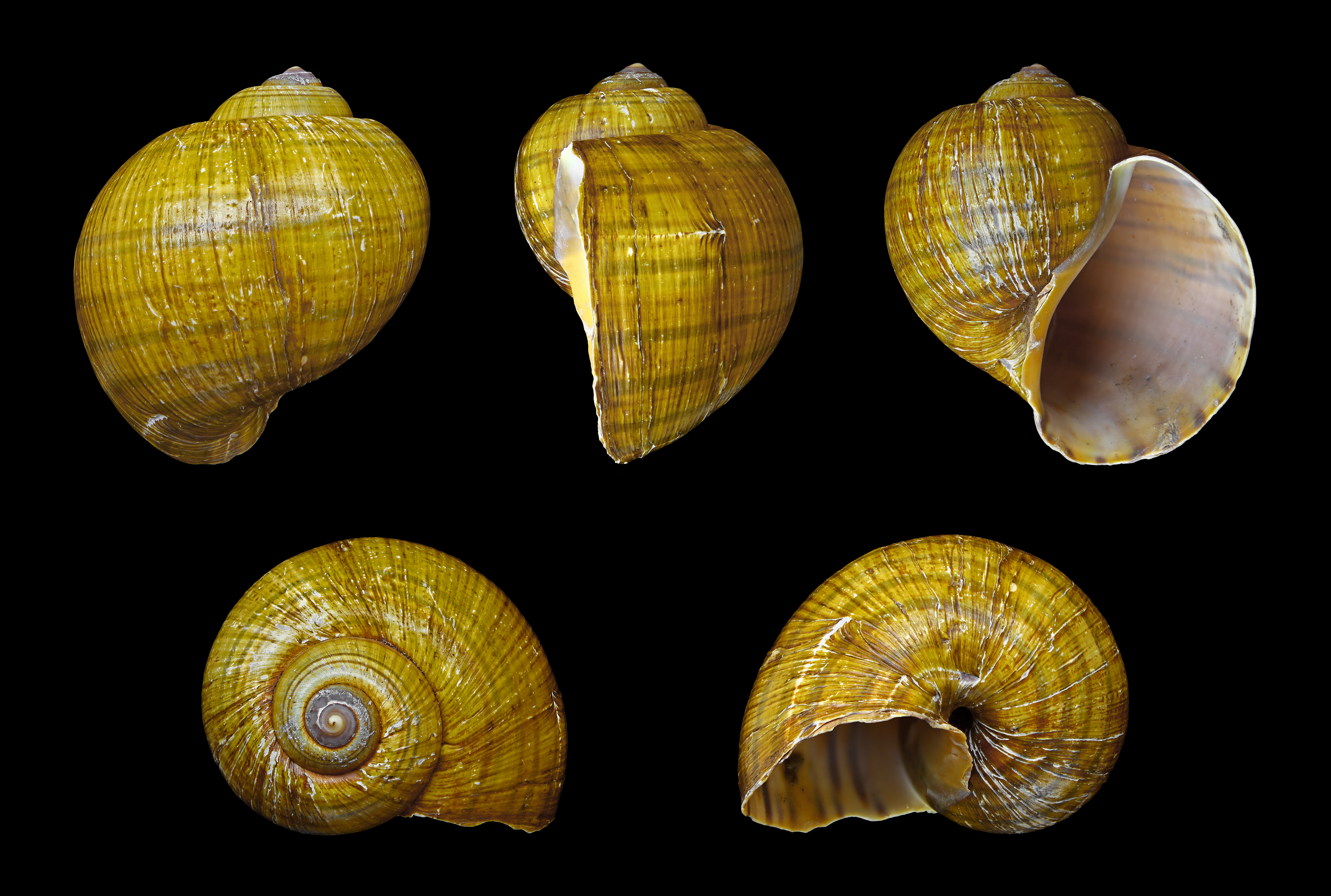
Figure 6.8: Pomacea canaliculata, H. Zell (own work)
Known Distribution of the Species
P. canaliculata is native from Argentina to the Amazon basin, as well as being introduced to most of southern, eastern, and south east Asia and the southern part of the United States.88 P. canaliculata is widely distributed in lakes, ponds and swamps throughout its native range of the Amazon Inferior Basin and the Plata Basin.89
Biogeographic Regions: nearctic (Introduced ); oriental (Introduced ); neotropical (Native); oceanic islands (Introduced ).90
First description
The French Biologist Jean Baptiste Lamarck first described P. canaliculata in the natural history of invertebrates in 1822.91
6.8.1 Scientific Research
Research categories
Much of the research on Pomacea canaliculata is categorised as Agronomy (7), Biotechnology and Applied Microbiology (4), Entomology (4), Environmental Sciences (4), Toxicology (4), Agriculture, Multidisciplinary (3).
Research summary
Much of the research on Pomacea canaliculata is concerned with ways to effectively control the invasive pest in agricultural settings, including the use of fish predators in rice fields, as well as molluscicides (De La Cruz and Joshi 2001; Sin 2006). Quinoa saponins a natural molluscicide were found to be an effective environmentally friendly alternative to synthetic molluscicides at eradicating Pomacea canaliculata, especially in direct seeded cultures of rice (Joshi et al. 2008). In a study in Thailand natural pathogens of Pomacea canaliculata were isolated from the soil - strains of Pseudomonas aeruginosa and P. fluoescens (Chobchuenchom and Bhumiratana 2003).
The human health risk posed by Pomacea canaliculata with regard to its transmission of parasites was the focus of several studies which monitored snail populations in aquatic bodies near populated areas. One study examined the effect of the building of a dam in central Laos PDR on numbers of all snails including Pomacea canaliculata - the intermediate host of A. cantonensis- it was the snail species found in the greatest numbers during 2010 and 2011; numbers increased greatly from 1.3% in 2010 to 53.3% in 2011 (Sri-aroon et al. 2015).
In 2006/7 tsunami and non-tsunami affected areas of Takua Pa District, Phang-Nga Province were examined for snails that transmit human parasitic diseases - 16 species were identified including Pomacea canaliculata. Knowledge of these medically important snails and their parasitic diseases, and prevention were given to Takua Pa people (Pusadee et al. 2010).
Another public health focus of the literature regarding Pomacea canaliculata concerned their potential use as biomonitors in waterways which could be subject to heavy metal pollution. This was shown to effect the tissues of the snails in an observable manner at levels below the safety limit, at a lake in Thailand (Dummee et al. 2012). Similarly another study focussed on the effect of copper sulphate exposure on the tissue of Pomacea canaliculata, they also found the snail to be an effective potential bioindicator of copper contamination in aquatic environments (Dummee et al. 2015).
A better understanding of the general biology of Pomacea canaliculata led some researchers to look at its life cycle (Arfan et al. 2015), whilst others examined genetic diversity (Shengzhang et al. 2011) and some specifically looked at tolerance of different climatic populations to dessication and cold (Wada and Matsukura 2011).
Differing cultivation methods were examined to note any effect on the mortality rate of rice due to Pomacea canaliculata infested ponds. One study noted that planting out 21 days after sowing gave a greater chance of survival for the rice seedlings (Horgan, Figueroa, and Almazan 2014).
Aquatic or marine? Although Pomacea canaliculata is largely a freshwater snail, reference is made to them occupying, marsh, swamp and marine habitats.92
Research locations
Snails in Beung Boraphet reservoir, Nakhon Sawan Province, central Thailand were examined in a study of metal pollution (Dummee et al. 2012). Areas of Takua Pa District, Phang-Nga Province were investigated for fresh- and brackish-water snails that transmit human parasitic diseases during 2006 and 2007 (Butraporn, P. 2010). Another study in Thailand looked at natural microbe pathogens in the soil (Chobchuenchom and Bhumiratana 2003).
The Philippines is the location of a couple of studies including one on rice cultivation methods to reduce mortality (Horgan, Figueroa, and Almazan 2014), and a molluscicide study in Munoz, Nueva Ecija (De La Cruz and Joshi 2001), as well as a genetic diversity analysis using a population from Los Banos (Shengzhang et al. 2011).
The effect of building a dam on the snail population was investigated at Khammouane Province, central Laos (Sri-aroon et al. 2015).
China (Yuyao, Taizhou, Fuzhou, Guangzhou, Nanning, Kunming) (Shengzhang et al. 2011), Japan (Kyushu and Luzon Mindanao) (Wada and Matsukura 2011) and Taiwan are also mentioned within the research literature (GH 1998).
6.8.2 Patent activity
There are 114 patent documents for Pomacea canaliculata, the most commonly cited patent document is for a modified plant chemical called ‘saponin’ that can be used to kill these molluscs in areas where they are considered a pest US20070196517A1.
Search the titles, abstracts and claims of patent documents for this species on the Lens or view the Lens public collection.
6.9 Vibrio harveyi
- Species name: Vibrio harveyi
- Kingdom: Bacteria
- Phylum: Proteobacteria GBIF record
Brief Description of the Species:
Vibrio harveyi, is a Gram-negative, bioluminescent, common marine bacterium in the same genus as Vibrio parahaemolyticus.93 V. harveyi is rod-shaped and can be found free-swimming in tropical marine waters, living commensally in the gut microflora of marine animals, a minority of V. harveyi strains are pathogenic to marine animals, including Gorgonian corals, oysters, prawns, lobsters, the common snook, barramundi, turbot, milkfish, and seahorses. It is responsible for luminous vibriosis, a disease that affects commercially farmed penaeid prawns such as P. monodon. Additionally, based on samples taken by ocean-going ships, V. harveyi is thought to be the cause of the milky seas effect, in which, during the night, a uniform blue glow is emitted from the seawater.94 Some glows can cover nearly 6,000 sq mi (16,000 km2).95
V. harveyi related strains have been isolated from the deep sea >1000m2, these may be ecologically differentiated strains, adapted to a different ecological niche or be distributed across the oceans (Hasan et al. 2015).
Although normally a benign bacteria in marine cultured environments, the prevalence of the pathogenic strains of V. harveyi - that differ by a few pathogenic determinant genes - can cause a problem in high nutrient, high density conditions leading to a rapid spread of virulent strains (Ben-Haim 2003).
Although closely related to the human pathogen V. parahaemolyticus, V. harveyi is not known as a human pathogen.96 This is one of the first organisms in which quorum sensing was described, whereby communities of bacteria communicate with each other via secreted signalling molecules synchronising community behaviour by regulating gene expression - both within and between bacteria species.97
Known Distribution of the Species
Vibrio harveyi is a common bacteria inhabiting largely tropical marine waters - as it requires sodium chloride, and prefers warmer water temperatures (Austin and Zhang 2006).
First description
Vibrio harveyi was named by Baumann et al. in 1981, further to the work of Johnson & Shunk, 1936 who originally named the species Achromobacter herveyi.98
6.9.1 Scientific Research
Research categories
Much of the research on Vibrio harveyi is categorised as Fisheries (72), Marine and Freshwater Biology (44), Veterinary Sciences (37), Immunology (30), Microbiology (24), Biotechnology and Applied Microbiology (23) and Biochemistry and Molecular Biology (19).
Research summary
The research effort on Vibrio harveyi is largely concerned with infection of fish, molluscs and crustaceans within the aquaculture industry. As such a lot of the V. harveyi literature is concerned with understanding the pathogenic strains of the bacteria that infects commercially farmed marine organisms, particularly shrimps: methods of rapidly detecting V. harveyi infections (Conejero and Hedreyda 2003), antimicrobial peptides produced by marine organisms that could combat V. harveyi and naturally occurring compounds that have antimicrobial properties against V. harveyi whilst being safe to cultured marine organisms (Ponprateep, Somboonwiwat, and Tassanakajon 2009; Maneechote et al. 2016). Shrimps infected with V. harveyi produce an antimicrobial peptide that has been shown to be effective in combating the bacteria when administered into a culture or injected into shrimps (Ponprateep, Somboonwiwat, and Tassanakajon 2009).
Overuse of antibiotics in aquaculture is blamed for an increase in antibiotic resistant strains of bacteria (Elmahdi, DaSilva, and Parveen 2016), so V. harveyi researchers are looking at alternative methods of combating the bacteria, including the use of novel natural compounds found in cyanobacteria (Maneechote et al. 2016) and extracts from Sargassum oligocystum (Nuestro et al. 2011).
Aquatic or marine?
Vibrio harveyi is found in marine and estuarine waters, however its presence has also been noted in Giant Freshwater Prawn larvae which inhabit brackish water as juveniles and freshwater as adults (Pande et al. 2013).
Research locations
Much of the research referencing specific locations for V. harveyi is from investigations into the cause of aquaculture shrimp mortalities as was the case in pond-cultured Penaeus monodon in the provinces of Bohol, Misamis Occidental, Lanao del Norte and Zamboanga del Sur, Philippines [Pena et al. (2003). Similarly dead shrimp larvae from hatcheries in Jepara, Indonesia (Prayitno and Latchford 1995), Southern Thailand (Ruangpan et al. 1999), and Iran (S et al. 2014) were found to contain V. harveyi. Seasonal changes in the composition of a variety of vibrio species including V. harveyi were investigated in samples taken from Yoshimi Bay, Hibiki-nada Sea, Japan.
6.9.2 Patent activity
Nucleic acid and amino acid sequences relating to pseudomonas aeruginosa for methods for the detection, prevention and treatment of pathological conditions resulting from bacterial infection including V. harveyi US6551795B1.
Microorganisms for therapy wherein the microorganisms gather in inflamed or cancerous tissues and cause cells to become leaky, resulting in production of antibodies, one such type of microorganism is attenuated Vibrio US20070212727A1
Search the titles, abstracts and claims of patent documents for this species on the Lens or view the Lens public collection.
6.10 Vibrio parahaemolyticus
- Species name: Vibrio parahaemolyticus
- Kingdom: Bacteria
- Phylum: Proteobacteria GBIF record
Brief Description of the Species:
Vibrio parahaemolyticus, is a gram negative bacteria that is found in estuarine, coastal and marine waters and is present in higher concentrations between May and October when temperatures are warmer (Letchumanan, Chan, and Lee 2014).99 V. parahaemolyticus responsible for the disease vibriosis in humans, contracted through eating raw contaminated shellfish such as oysters, or exposure of a wound to infected salt or brackish water.100
V. parahaemolyticus is found in a free swimming state, using a single flagellum to attach to zooplankton, fish, shellfish or suspended matter in the water (Letchumanan, Chan, and Lee 2014).
The prevalence of the pathogenic strains of V. parahaemolyticus isolated from seafood, and clinical samples shows it to have a worldwide distribution across South East Asia, Europe, and US posing an ongoing health threat to the population. Within Asia, samples have been found in seafood in markets in China, Malaysia, India, Bangladesh, Taiwan, Laos, Hong Kong and Japan (Letchumanan, Chan, and Lee 2014).
Known Distribution of the Species
“Present in the Gulf of Mexico” GBIF record.
First description
Vibrio parahaemolyticus was named by Sazaki et al.in 1963, further to the work of Fujino et al. in 1951 who originally named the species Pasteurella parahaemolytica.101.
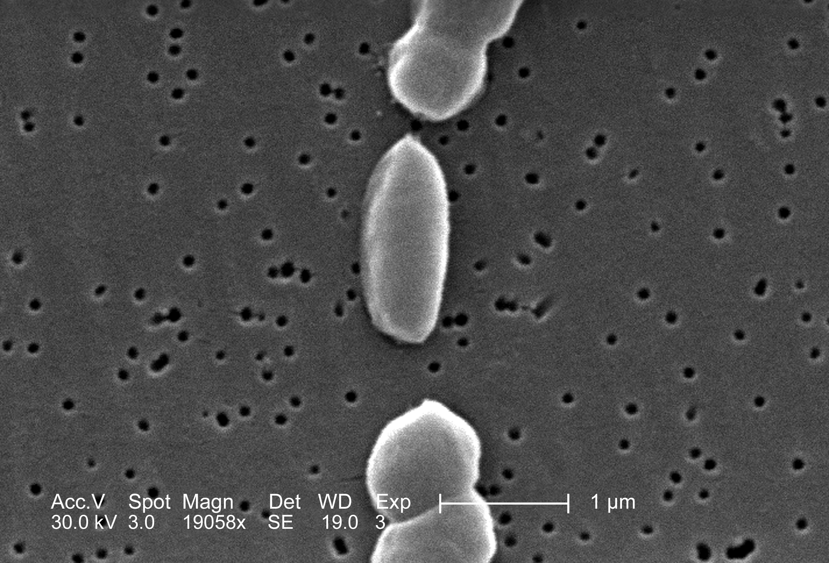
Figure 6.9: Vibrio parahaemolyticus, Janice Carr/CDC
6.10.1 Scientific Research
Research categories
Much of the research on Vibrio parahaemolyticus is categorised as Microbiology (60), Food Science and Technology (30), Biotechnology and Applied Microbiology (29), Fisheries (29), Veterinary Sciences (21), Infectious diseases (19), Marine and Freshwater Biology (17), Immunology (11) and so on.
Research summary
The research effort on Vibrio parahaemolyticus is largely concerned with infection of fish, molluscs and crustaceans within the aquaculture industry, as well as infection of humans that eat infected seafood. As such a lot of the V. parahaemolyticus literature is concerned with understanding the pathogenic strains of the bacteria that infects humans and commercially reared seafood including: methods of rapidly detecting strains causing an infection , vaccines offering protection and ways of treating the infection (Rahman et al. 2006; Hu and Sun 2011). Shrimps infected with Vibrio parahaemolyticus can develop acute hepatopancreatic necrosis disease causing severe mortalities in SE Asia and Mexico, some research has been directed to noting any genetic difference between bacterial infections from different geographical areas.
Overuse of antibiotics in aquaculture is blamed for an increase in antibiotic resistant strains of Vibrio parahaemolyticus, so researchers are also looking at alternative methods of combating the bacteria: including the use of bacteria phage - such as one discovered in Laos (Elmahdi, DaSilva, and Parveen 2016; Noboru et al. 1999). In ensuring people don’t become sick from eating seafood, some research has been focused on food preservatives that extend the life of seafood because of their inhibitory effect on Vibrio parahaemolyticussuch as botanical extracts from Terminalia chebula (P, Chitsiri, and Nophadon 2012) and Zingiber spectabile, and best practice for safe processing of seafood (Sivasothy et al. 2013; Boonyawantang et al. 2012). There are many studies noting the range of strains present in certain geographic areas, in Taiwan and 14 other countries 535 strains were identified of Vibrio parahaemolyticus - either by testing samples of infected faeces of people from clinical settings, or sampling water and noting the strains of Vibrio parahaemolyticus present (Wong et al. 2007; Wootipoom et al. 2007). Certain strains are given more attention, as they are more virulent, pathogenic and novel - so there is little resistance in the local human population - it is these strains, such as the pandemic strain, that are of more concern in the literature.
Aquatic or marine?
Vibrio parahaemolyticus is found in marine and brackish waters, however its presence has also been noted in a tropical coastal lagoon from 2 samples collected in the estuary near Yor Island, Songkhla Lake, Thailand (Thongchankaew et al. 2011).
Research locations
Much of the research involving samples taken from humans infected with Vibrio parahaemolyticus reference a country for example:US, Italy, Brazil, Philippines, Malaysia, China, India, Thailand, Iran, South Africa and Australia (Elmahdi, DaSilva, and Parveen 2016). Some clinical studies are more specific in noting a hospital or region: a study conducted at Hat Yai Hospital in southern Thailand (Wootipoom et al. 2007) for example, and a similar stool sample study in the infectious Diseases Hospital in Kolkata, India examining strains present in infected samples using the PFGE analysis technique to identify the strains (Pazhani et al. 2014). Food poisoning cases in Kangawa Prefecture, Japan were the source of 18 strains of V. parahaemolyticus studied in laboratories in Thailand to note the specific genes present (Okitsu et al. 1997).
Forty two strains of Vibrio parahaemolyticus were isolated form samples of water taken form the estuaries around Bangladesh in the Bay of Bengal, with some being of the virulent and pathogenic strains posing a risk to inhabitants of coastal villages in the area (Alam et al. 2009). A study on patients with diarrhoea in North Jakarta, Indonesia, found Vibrio parahaemolyticus was responsible for a substantial number of cases, with incidence being highest in the dry season (June and July), many of the samples showed antibiotic resistance to some antibiotics (Lesmana et al. 2001).
Samples of seafood were analysed from Hong Kong, Indonesia, Thailand and Vietnam - of the samples taken 45.9% contained Vibrio parahaemolyticus, with more of the samples from Hong Kong and Thailand containing the bacteria (Wong et al. 1999).
170 strains of V. parahaemolyticus were isolated from water samples collected along the Georgian coast of the Black Sea, water temperature was shown to be a factor in its presence especially in the Green cape samples (Haley et al. 2014).
6.10.2 Patent activity
The top cited patent document involving V. parahaemolyticus is for identification of essential genes used to help V. parahaemolyticus multiply US20040029129A1 - a sequence of nucleic acids is used to understand which are the key important proteins in the bacteria and then drugs, or antibodies can be produced to prevent replication of the bacteria.
The patent document with the largest family is for the use of a peptide as a therapeutic agent WO2009043477A2 when someone has a V. parahaemolyticus infection.
Search the titles, abstracts and claims of patent documents for this species on the Lens or view the Lens public collection.
References
Alam, M., W. B. Chowdhury, N. A. Bhuiyan, A. Islam, N. A. Hasan, G. B. Nair, H. Watanabe, et al. 2009. “Serogroup, Virulence, and Genetic Traits of Vibrio Parahaemolyticus in the Estuarine Ecosystem of Bangladesh.” Applied and Environmental Microbiology 75 (19). American Society for Microbiology: 6268–74. https://doi.org/10.1128/aem.00266-09.
Ali, Ahyaudin B. 1993. “Aspects of the Fecundity of the Feral Catfish, Clarias Macrocephalus (Gunther), Population Obtained from the Rice Fields Used for Rice-Fish Farming, in Malaysia.” Hydrobiologia 254 (2). Springer Nature: 81–89. https://doi.org/10.1007/bf00014311.
Alongi, D.M, V.C Chong, P Dixon, A Sasekumar, and F Tirendi. 2003. “The Influence of Fish Cage Aquaculture on Pelagic Carbon Flow and Water Chemistry in Tidally Dominated Mangrove Estuaries of Peninsular Malaysia.” Marine Environmental Research 55 (4). Elsevier BV: 313–33. https://doi.org/10.1016/s0141-1136(02)00276-3.
Arfan, Gilal, Muhamad Rita, Omar Dzolkifli, Abd Aziz Nor, and Gnanasegaram Manjeri. 2015. “Comparative Life Cycle Studies of Pomacea Maculata and Pomacea Canaliculata on Rice (Oryza Sativa).” Pakistan Journal of Agricultural Sciences 52 (December): 1079–83.
Aungsuchawan, Sirinda, Amy O. Ball, Robert W. Chapman, Eleanor Shepard, Craig L. Browdy, and Boonsirm Withyachumnarnkul. 2008. “Evaluation of Published Microsatellites for Paternity Analysis in the Pacific White Shrimp Litopenaeus Vannamei.” ScienceAsia 34 (1). Science Society of Thailand - Mahidol University: 115. https://doi.org/10.2306/scienceasia1513-1874.2008.34.115.
Austin, B., and X-H. Zhang. 2006. “Vibrio Harveyi: A Significant Pathogen of Marine Vertebrates and Invertebrates.” Letters in Applied Microbiology 43 (2). Wiley: 119–24. https://doi.org/10.1111/j.1472-765x.2006.01989.x.
Bahari, A, M Afsharnasab, A Motalbei Moghanjoghi, G Azaritakami, and M and Shrifrohani. 2015. “Experimental Pathogenicity of Shrimp, Penaeus Vannamei Exposed to Monodon Baculovirus (Mbv).” Iranian Journal of Fisheries Sciences 14 (2). http://jifro.ir/article-1-1888-en.html.
Baharom, Zarith Sufiani, and Mohd Yusoff Ishak. 2015. “Determination of Heavy Metal Accumulation in Fish Species in Galas River, Kelantan and Beranang Mining Pool, Selangor.” Procedia Environmental Sciences 30. Elsevier BV: 320–25. https://doi.org/10.1016/j.proenv.2015.10.057.
Ben-Haim, Y. 2003. “Vibrio Coralliilyticus Sp. Nov., a Temperature-Dependent Pathogen of the Coral Pocillopora Damicornis.” International Journal of Systematic and Evolutionary Microbiology 53 (1). Microbiology Society: 309–15. https://doi.org/10.1099/ijs.0.02402-0.
Bentsen, Hans B., Ambekar E. Eknath, Marietta S. Palada-de Vera, Jodecel C. Danting, Hernando L. Bolivar, Ruben A. Reyes, Edna E. Dionisio, et al. 1998. “Genetic Improvement of Farmed Tilapias: Growth Performance in a Complete Diallel Cross Experiment with Eight Strains of Oreochromis Niloticus.” Aquaculture 160 (1-2). Elsevier BV: 145–73. https://doi.org/10.1016/s0044-8486(97)00230-5.
Biju, N, G Sathiyaraj, M Raj, V Shanmugam, B Baskaran, U Govindan, G Kumaresan, KK Kasthuriraju, and TSRY Chellamma. 2016. “High Prevalence of Enterocytozoon Hepatopenaei in Shrimps Penaeus Monodon and Litopenaeus Vannamei Sampled from Slow Growth Ponds in India.” Diseases of Aquatic Organisms 120 (3). Inter-Research Science Center: 225–30. https://doi.org/10.3354/dao03036.
Bombeo, Ruby F, Armando C Fermin, and Josefa D Tan-Fermin. 2002. “Nursery Rearing of the Asian Catfish, Clarias Macrocephalus (Gunther), at Different Stocking Densities in Cages Suspended in Tanks and Ponds.” Aquaculture Research 33 (13). Wiley: 1031–6. https://doi.org/10.1046/j.1365-2109.2002.00763.x.
Bonami, Jean-Robert, and Joannes Sri Widada. 2011. “Viral Diseases of the Giant Fresh Water Prawn Macrobrachium Rosenbergii: A Review.” Journal of Invertebrate Pathology 106 (1). Elsevier BV: 131–42. https://doi.org/10.1016/j.jip.2010.09.007.
Boonyawantang, Arisara, Warapa Mahakarnchanakul, Chitsiri Rachtanapun, and Waraporn Boonsupthip. 2012. “Behavior of Pathogenic Vibrio Parahaemolyticus in Prawn in Response to Temperature in Laboratory and Factory.” Food Control 26 (2). Elsevier BV: 479–85. https://doi.org/10.1016/j.foodcont.2012.02.009.
Caipang, Christopher Marlowe A., and Mary Paz N. Aguana. 2010. “Rapid Diagnosis of Vibriosis and White Spot Syndrome (WSS) in the Culture of Shrimp, Penaeus Monodon in Philippines.” Veterinary Research Communications 34 (7). Springer Nature: 597–605. https://doi.org/10.1007/s11259-010-9434-x.
Chaivisuthangkura, P, T Tejangkura, S Rukpratanporn, S Longyant, W Sithigorngul, and P Sithigorngul. 2006. “Polyclonal Antibodies Specific for VP1 and VP3 Capsid Proteins of Taura Syndrome Virus (TSV) Produced via Gene Cloning and Expression.” Diseases of Aquatic Organisms 69 (April). Inter-Research Science Center: 249–53. https://doi.org/10.3354/dao069249.
Chaweepack, Tidaporn, Surachart Chaweepack, Boonyee Muenthaisong, Lila Ruangpan, Kei Nagata, and Kaeko Kamei. 2014. “Effect of Galangal (Alpinia Galanga Linn.) Extract on the Expression of Immune-Related Genes and Vibrio Harveyi Resistance in Pacific White Shrimp (Litopenaeus Vannamei).” Aquaculture International 23 (1). Springer Nature: 385–99. https://doi.org/10.1007/s10499-014-9822-2.
Chobchuenchom, Wimol, and Amaret Bhumiratana. 2003. “Isolation and Characterization of Pathogens Attacking Pomacea Canaliculata.” World Journal of Microbiology and Biotechnology 19 (9). Springer Nature: 903–6. https://doi.org/10.1023/b:wibi.0000007312.97058.48.
Choo, Jenny, Nuraini Binti Mohd Sabri, Daniel Tan, Aazani Mujahid, and Moritz Müller. 2015. “Heavy Metal Resistant Endophytic Fungi Isolated from Nypa Fruticans in Kuching Wetland National Park.” Ocean Science Journal 50 (2). Springer Nature: 445–53. https://doi.org/10.1007/s12601-015-0040-2.
Claydon, Kerry, Rahimah Awg Haji Tahir, Hajijah Mohd Said, Mahani Haji Lakim, and Wanidawati Tamat. 2010. “Prevalence of Shrimp Viruses in Wild Penaeus Monodon from Brunei Darussalam.” Aquaculture 308 (3-4). Elsevier BV: 71–74. https://doi.org/10.1016/j.aquaculture.2010.08.015.
Conejero, M.J.U., and C.T. Hedreyda. 2003. “Isolation of Partial toxR Gene of Vibrio Harveyi and Design of toxR-Targeted PCR Primers for Species Detection.” Journal of Applied Microbiology 95 (3). Wiley: 602–11. https://doi.org/10.1046/j.1365-2672.2003.02020.x.
Cruz CPP, and Paller VGV. 2012. “Occurrence of Neoechinorhynchus Sp (Acanthocephala: Neoechinorhynchidae) in Cultured Tilapia,[Oreochromis Niloticus (L.), Perciformes: Ciclidae] from Sampaloc Lake, Philippines.” Asia Life Sciences 21 (1): 287–98.
Davis, T.L.O. 1986. “Migration Patterns in Barramundi, Lates Calcarifer (Bloch), in van Diemen Gulf, Australia, with Estimates of Fishing Mortality in Specific Areas.” Fisheries Research 4 (3-4). Elsevier BV: 243–58. https://doi.org/10.1016/0165-7836(86)90006-8.
De La Cruz, M.S., and R.C. Joshi. 2001. “Efficacy of Commercial Molluscicide Formulations Against the Golden Apple Snail Pomacea Canaliculata (Lamarck)” 84 (March): 51–55.
Dummee, Vipawee, Maleeya Kruatrachue, Wachareeporn Trinachartvanit, Phanwimol Tanhan, Prayad Pokethitiyook, and Praneet Damrongphol. 2012. “Bioaccumulation of Heavy Metals in Water, Sediments, Aquatic Plant and Histopathological Effects on the Golden Apple Snail in Beung Boraphet Reservoir, Thailand.” Ecotoxicology and Environmental Safety 86 (December). Elsevier BV: 204–12. https://doi.org/10.1016/j.ecoenv.2012.09.018.
Dummee, Vipawee, Phanwimol Tanhan, Maleeya Kruatrachue, Praneet Damrongphol, and Prayad Pokethitiyook. 2015. “Histopathological Changes in Snail, Pomacea Canaliculata, Exposed to Sub-Lethal Copper Sulfate Concentrations.” Ecotoxicology and Environmental Safety 122 (December). Elsevier BV: 290–95. https://doi.org/10.1016/j.ecoenv.2015.08.010.
Duponchelle, Fabrice, Philippe Cecchi, Daniel Corbin, Jesus Nuñez, and Marc Legendre. 1999. Environmental Biology of Fishes 56 (4). Springer Nature: 375–87. https://doi.org/10.1023/a:1007588010824.
Elmahdi, Sara, Ligia V. DaSilva, and Salina Parveen. 2016. “Antibiotic Resistance of Vibrio Parahaemolyticus and Vibrio Vulnificus in Various Countries: A Review.” Food Microbiology 57 (August). Elsevier BV: 128–34. https://doi.org/10.1016/j.fm.2016.02.008.
Engle, Carole R., Aaron McNevin, Phoebe Racine, Claude E. Boyd, Duangchai Paungkaew, Rawee Viriyatum, Huynh Quoc Tinh, and Hang Ngo Minh. 2017. “Economics of Sustainable Intensification of Aquaculture: Evidence from Shrimp Farms in Vietnam and Thailand.” Journal of the World Aquaculture Society 48 (2). Wiley: 227–39. https://doi.org/10.1111/jwas.12423.
Fermin, Armando C., Ma. Edna C. Bolivar, and Albert Gaitan. 1996. “Nursery Rearing of the Asian Sea Bass,Lates Calcarifer, Fry in Illuminated Floating Net Cages with Different Feeding Regimes and Stocking Densities.” Aquatic Living Resources 9 (1). EDP Sciences: 43–49. https://doi.org/10.1051/alr:1996006.
G, Wang, Tan S, Li S, and Ye Haihui. 2008. “Genetic Diversity and Differentiation of Three Populations of Penaeus Monodon Fabricus” 27 (January): 113–21.
GH, Baker. 1998. The Golden Apple Snail, Pomacea Canaliculata (Lamarck) (Mollusca: Ampullariidae), a Potential Invader of Fresh Water Habitats in Australia. Vol. 2. University of Queensland.
Gibson-Kueh, S, D Chee, J Chen, Y H Wang, S Tay, L N Leong, M L Ng, J B Jones, P K Nicholls, and H W Ferguson. 2011. “The Pathology of ‘Scale Drop Syndrome’ in Asian Seabass, Lates Calcarifer Bloch, a First Description.” Journal of Fish Diseases 35 (1). Wiley: 19–27. https://doi.org/10.1111/j.1365-2761.2011.01319.x.
Glenn, K. L., L. Grapes, T. Suwanasopee, D. L. Harris, Y. Li, K. Wilson, and M. F. Rothschild. 2005. “SNP Analysis ofAMY2andCTSLgenes inLitopenaeus vannameiandPenaeus Monodonshrimp.” Animal Genetics 36 (3). Wiley: 235–36. https://doi.org/10.1111/j.1365-2052.2005.01274.x.
Guerrero, Rafael D. 2008. “Tilapia Farming: A Global Review (1924-2004).” Asia Life SCiences 17 (2): 207–29.
Ha, Tran Thi Phung, Han van Dijk, Roel Bosma, and Le Xuan Sinh. 2013. “LIVELIHOOD CAPABILITIES AND PATHWAYS OF SHRIMP FARMERS IN THE MEKONG DELTA, VIETNAM.” Aquaculture Economics & Management 17 (1). Informa UK Limited: 1–30. https://doi.org/10.1080/13657305.2013.747224.
Haley, Bradd J., Tamar Kokashvili, Ana Tskshvediani, Nino Janelidze, Nino Mitaishvili, Christopher J. Grim, Guillaume Constantin de Magny, et al. 2014. “Molecular Diversity and Predictability of Vibrio Parahaemolyticus Along the Georgian Coastal Zone of the Black Sea.” Frontiers in Microbiology 5. Frontiers Media SA. https://doi.org/10.3389/fmicb.2014.00045.
Hasan, Nur A., Christopher J. Grim, Erin K. Lipp, Irma N. G. Rivera, Jongsik Chun, Bradd J. Haley, Elisa Taviani, et al. 2015. “Deep-Sea Hydrothermal Vent Bacteria Related to Human pathogenicVibriospecies.” Proceedings of the National Academy of Sciences 112 (21). Proceedings of the National Academy of Sciences: E2813–E2819. https://doi.org/10.1073/pnas.1503928112.
Hien, Tran Thi Thanh, Tran Ngoc HAI, Nguyen Thanh PHUONG, Hiroshi Y. OGATA, and Marcy N. WILDER. 2005. “The Effects of Dietary Lipid Sources and Lecithin on the Production of Giant Freshwater Prawn Macrobrachium Rosenbergii Larvae in the Mekong Delta Region of Vietnam.” Fisheries Science 71 (2). Springer Nature: 279–86. https://doi.org/10.1111/j.1444-2906.2005.00961.x.
Hinrichs, Saskia, Inga Nordhaus, and Simon Joscha Geist. 2008. “Status, Diversity and Distribution Patterns of Mangrove Vegetation in the Segara Anakan Lagoon, Java, Indonesia.” Regional Environmental Change 9 (4). Springer Nature: 275–89. https://doi.org/10.1007/s10113-008-0074-4.
Ho, Teerapong, Pratchayapong Yasri, Sakol Panyim, and Apinunt Udomkit. 2011. “Double-Stranded RNA Confers Both Preventive and Therapeutic Effects Against Penaeus Stylirostris Densovirus (PstDNV) in Litopenaeus Vannamei.” Virus Research 155 (1). Elsevier BV: 131–36. https://doi.org/10.1016/j.virusres.2010.09.009.
Horgan, Finbarr G., Jesús Yanes Figueroa, and Maria Liberty P. Almazan. 2014. “Seedling Broadcasting as a Potential Method to Reduce Apple Snail Damage to Rice.” Crop Protection 64 (October). Elsevier BV: 168–76. https://doi.org/10.1016/j.cropro.2014.06.022.
Hu, Yong-hua, and Li Sun. 2011. “A Bivalent Vibrio Harveyi DNA Vaccine Induces Strong Protection in Japanese Flounder (Paralichthys Olivaceus).” Vaccine 29 (26). Elsevier BV: 4328–33. https://doi.org/10.1016/j.vaccine.2011.04.021.
I, Kaswani, Kamaruzaman J, and Nurun-Nadhuzah M I. 2007. “A Study of Forest Structure, Diversity Index and Above-Ground Biomass at Tok Bali Mangrove Forest, Kelantan, Malaysia.” 5th WSEAS Int. Conf. On Environment, Ecosystems and Development, Tenerife, Spain. World Scientific; Engineering Acad; Soc. http://www.worldses.org/journals/environment/environment-february2007.htm.
JA, Mantiquilla, Solano JDM, Abad RG, Rivero GC, and Silvosa CSC. 2015. “Callus Induction from Young Leaf Explants of Nipa (Nypa Fruticans Wurmb.).” Asia Life Sciences 24 (1). Asia Life Sciences: 409–26.
Jantrakajorn, S., and J. Wongtavatchai. 2016. “Egg Surface Decontamination with Bronopol Increases Larval Survival of Nile Tilapia, Oreochromis Niloticus.” Czech Journal of Animal Science 60 (No. 10). Czech Academy of Agricultural Sciences: 436–42. https://doi.org/10.17221/8523-cjas.
Jaree, Phattarunda, Anchalee Tassanakajon, and Kunlaya Somboonwiwat. 2012. “Effect of the Anti-Lipopolysaccharide Factor Isoform 3 (ALFPm3) from Penaeus Monodon on Vibrio Harveyi Cells.” Developmental & Comparative Immunology 38 (4). Elsevier BV: 554–60. https://doi.org/10.1016/j.dci.2012.09.001.
Jian, Shuguang, Jiawei Ban, Hai Ren, and Haifei Yan. 2010. “Low Genetic Variation Detected Within the Widespread Mangrove Species Nypa Fruticans (Palmae) from Southeast Asia.” Aquatic Botany 92 (1). Elsevier BV: 23–27. https://doi.org/10.1016/j.aquabot.2009.09.003.
Joshi, Ravindra C., Ricardo San Martı'n, Cesar Saez-Navarrete, John Alarcon, Javier Sainz, Mina M. Antolin, Antonio R. Martin, and Leocadio S. Sebastian. 2008. “Efficacy of Quinoa (Chenopodium Quinoa) Saponins Against Golden Apple Snail (Pomacea Canaliculata) in the Philippines Under Laboratory Conditions.” Crop Protection 27 (3-5). Elsevier BV: 553–57. https://doi.org/10.1016/j.cropro.2007.08.010.
Jr, Rasco Eufemio T. 2010. “Biology of Nipa Palm (Nypa Fruticans Wurmb., Arecaceae) and Its Potential for Alcohol Production.” Asia Life Sciences 19 (2). Asia Life Sciences: 373–88.
Jung, Hyungtaek, Byung-Ha Yoon, Woo-Jin Kim, Dong-Wook Kim, David Hurwood, Russell Lyons, Krishna Salin, et al. 2016. “Optimizing Hybrid de Novo Transcriptome Assembly and Extending Genomic Resources for Giant Freshwater Prawns (Macrobrachium Rosenbergii): The Identification of Genes and Markers Associated with Reproduction.” International Journal of Molecular Sciences 17 (5). MDPI AG: 690. https://doi.org/10.3390/ijms17050690.
Jusoh, Wan Faridah Akmal Wan, Nor Rasidah Hashim, and Zelina Z. Ibrahim. 2010. “Firefly Distribution and Abundance on Mangrove Vegetation Assemblages in Sepetang Estuary, Peninsular Malaysia.” Wetlands Ecology and Management 18 (3). Springer Nature: 367–73. https://doi.org/10.1007/s11273-009-9172-4.
Jutagate, Tuantong, Achara Rattanachai, Suriya Udduang, Sithan Lek-Ang, and Sovan Lek. 2016. “Spatio-Temporal Variations in Abundance and Assemblage Patterns of Fish Larvae and Their Relationships to Environmental Variables in Sirindhron Reservoir of the Lower Mekong Basin, Thailand.” Indian Journal of Fisheries 63 (3). Central Marine Fisheries Research Institute, Kochi. https://doi.org/10.21077/ijf.2016.63.3.54491-02.
Kaew-on, Saichai, Nontawith Areechon, and Penkhae Wanchaitanawong. 2016. “Effects of Pediococcus Pentosaceus Pkwa-1 and Bacillus Subtilis Ba04 on Growth Performances, Immune Responses and Disease Resistance Against Aeromonas Hydrophila in Nile Tilapia (Oreochromis Niloticus Linn.).” Chiang Mai Journal of Science 43 (5). Chiang Mai University: 997–1006. http://epg.science.cmu.ac.th/ejournal/.
Kamal, Abu Hena Md. Mostofa, and Graham C. Mair. 2005. “Salinity Tolerance in Superior Genotypes of Tilapia, Oreochromis Niloticus, Oreochromis Mossambicus and Their Hybrids.” Aquaculture 247 (1-4). Elsevier BV: 189–201. https://doi.org/10.1016/j.aquaculture.2005.02.008.
Kangpanich, Chanpim, Jarunan Pratoomyot, Nisa Siranonthana, and Wansuk Senanan. 2016. “Effects of Arachidonic Acid Supplementation in Maturation Diet on Female Reproductive Performance and Larval Quality of Giant River Prawn (Macrobrachium Rosenbergii).” PeerJ 4 (November). PeerJ: e2735. https://doi.org/10.7717/peerj.2735.
Kanjanaworakul, Poonmanee, Prapansak Srisapoome, Orathai Sawatdichaikul, and Supawadee Poompuang. 2014. “cDNA Structure and the Effect of Fasting on Myostatin Expression in Walking Catfish (Clarias Macrocephalus, Günther 1864).” Fish Physiology and Biochemistry 41 (1). Springer Nature: 177–91. https://doi.org/10.1007/s10695-014-0015-8.
Karaket, Thuchapol, and Supawadee Poompuang. 2012. “CERVUS Vs. COLONY for Successful Parentage and Sibship Determinations in Freshwater Prawn Macrobrachium Rosenbergii de Man.” Aquaculture 324-325 (January). Elsevier BV: 307–11. https://doi.org/10.1016/j.aquaculture.2011.10.045.
Karapanagiotidis, Ioannis T., Michael V. Bell, David C. Little, and Amararatne Yakupitiyage. 2007. “Replacement of Dietary Fish Oils by Alpha-Linolenic Acid-Rich Oils Lowers Omega 3 Content in Tilapia Flesh.” Lipids 42 (6). Wiley: 547–59. https://doi.org/10.1007/s11745-007-3057-1.
KD, Hyde, and Sarma VV. 2006. “Biodiversity and Ecological Observations on Filamentous Fungi of Mangrove Palm Nypa Fruticans Wurumb (Liliopsida-Arecales) Along the Tutong River, Brunei.” INDIAN JOURNAL OF MARINE SCIENCES 35 (4). NISCAIR: 297. http://nopr.niscair.res.in/handle/123456789/1529.
Khimmakthong, Umaporn, Panchalika Deachamag, Amornrat Phongdara, and Wilaiwan Chotigeat. 2011. “Stimulating the Immune Response of Litopenaeus Vannamei Using the Phagocytosis Activating Protein (PAP) Gene.” Fish & Shellfish Immunology 31 (3). Elsevier BV: 415–22. https://doi.org/10.1016/j.fsi.2011.06.010.
Koolboon, Urai, Skorn Koonawootrittriron, Wongpathom Kamolrat, and Uthairat Na-Nakorn. 2014. “Effects of Parental Strains and Heterosis of the Hybrid Between Clarias Macrocephalus and Clarias Gariepinus.” Aquaculture 424-425 (March). Elsevier BV: 131–39. https://doi.org/10.1016/j.aquaculture.2013.12.023.
Koolkalya, Sontaya, Amonsak Sawusdee, and Jutagate Tuantong. 2015. “Chronicle of Marine Fisheries in the Gulf of Thailand: Variations, Trends and Patterns.” Indian Journal of Geo-Marine Sciences 44 (9). NISCAIR: 1302–9. https://www.researchgate.net/publication/305543157_Chronicle_of_marine_fisheries_in_the_Gulf_of_Thailand_Variations_trends_and_patterns.
Kosai, Piya, Wannee Jiraungkoorskul, Tawan Thammasunthorn, and Kanitta Jiraungkoorskul. 2009. “Reduction of Copper-Induced Histopathological Alterations by Calcium Exposure in Nile Tilapia (Oreochromis Niloticus).” Toxicology Mechanisms and Methods 19 (6-7). Informa UK Limited: 461–67. https://doi.org/10.1080/15376510903173674.
Kuedo, Zulkiflee, Anantita Sangsuriyawong, Wanwimol Klaypradit, Varomyalin Tipmanee, and Pennapa Chonpathompikunlert. 2016. “Effects of Astaxanthin from Litopenaeus Vannamei on Carrageenan-Induced Edema and Pain Behavior in Mice.” Molecules 21 (3). MDPI AG: 382. https://doi.org/10.3390/molecules21030382.
Lesmana, Murad, Decy Subekti, Cyrus H Simanjuntak, Periska Tjaniadi, James R Campbell, and Buhari A Oyofo. 2001. “Vibrio Parahaemolyticus Associated with Cholera-Like Diarrhea Among Patients in North Jakarta, Indonesia.” Diagnostic Microbiology and Infectious Disease 39 (2). Elsevier BV: 71–75. https://doi.org/10.1016/s0732-8893(00)00232-7.
Letchumanan, Vengadesh, Kok-Gan Chan, and Learn-Han Lee. 2014. “Vibrio Parahaemolyticus: A Review on the Pathogenesis, Prevalence, and Advance Molecular Identification Techniques.” Frontiers in Microbiology 5 (December). Frontiers Media SA. https://doi.org/10.3389/fmicb.2014.00705.
Liao, I Chiu, and Yew-Hu Chien. 2011. “The Pacific White Shrimp, Litopenaeus Vannamei, in Asia: The World’s Most Widely Cultured Alien Crustacean.” In In the Wrong Place - Alien Marine Crustaceans: Distribution, Biology and Impacts, 489–519. Springer Netherlands. https://doi.org/10.1007/978-94-007-0591-3_17.
Mair, G C, J S Abucay, T A Abella, J A Beardmore, and DOF Skibinski. 1997. “Genetic Manipulation of Sex Ratio for the Large-Scale Production of All-Male Tilapia Oreochromis Niloticus.” Canadian Journal of Fisheries and Aquatic Sciences 54 (2). Canadian Science Publishing: 396–404. https://doi.org/10.1139/f96-282.
Maneechote, Nipat, Boon-ek Yingyongnarongkul, Apichart Suksamran, and Saisamorn Lumyong. 2016. “Inhibition ofVibriospp. By 2-Hydroxyethyl-11-Hydroxyhexadec-9-Enoate of Marine CyanobacteriumLeptolyngbyasp. LT19.” Aquaculture Research 48 (5). Wiley: 2088–95. https://doi.org/10.1111/are.13043.
Marcussen, Helle, Peter E. Holm, Le Thai Ha, and Anders Dalsgaard. 2007. “Food Safety Aspects of Toxic Element Accumulation in Fish from Wastewater-Fed Ponds in Hanoi, Vietnam.” Tropical Medicine & International Health 12 (November). Wiley: 34–39. https://doi.org/10.1111/j.1365-3156.2007.01939.x.
Mohd-Yusof, N. Y., O. Monroig, A. Mohd-Adnan, K.-L. Wan, and D. R. Tocher. 2010. “Investigation of Highly Unsaturated Fatty Acid Metabolism in the Asian Sea Bass, Lates Calcarifer.” Fish Physiology and Biochemistry 36 (4). Springer Nature: 827–43. https://doi.org/10.1007/s10695-010-9409-4.
Mohr, PG, NJG Moody, J Hoad, LM Williams, RO Bowater, DM Cummins, JA Cowley, and StJ Crane M. 2015. “New Yellow Head Virus Genotype (YHV7) in Giant Tiger Shrimp Penaeus Monodon Indigenous to Northern Australia.” Diseases of Aquatic Organisms 115 (3). Inter-Research Science Center: 263–68. https://doi.org/10.3354/dao02894.
Moore, R. 1982. “Spawning and Early Life History of Burramundi, Lates Calcarifer (Bloch), in Papua New Guinea.” Marine and Freshwater Research 33 (4). CSIRO Publishing: 647. https://doi.org/10.1071/mf9820647.
Mosha, Erastus John, and Wenresti Glino Gallardo. 2013. “Distribution and Size Composition of Penaeid Prawns, Penaeus Monodon and Penaeus Indicus in Saadan Estuarine Area, Tanzania.” Ocean & Coastal Management 82 (September). Elsevier BV: 51–63. https://doi.org/10.1016/j.ocecoaman.2013.05.003.
MZ, Khaironizam, and Norma-Rashid Y. 2003. “First Record of the Mudskipper, Periophthalmodon Septemradiatus (Hamilton) (Teleostei : Gobiidae) from Peninsular Malaysia.” Raffles Bulletin of Zoology 51 (1). National University of Singapore: 97–100. https://archive.org/details/raffles-bulletin-zoology-51-097-100/page/n0.
Na-Nakorn, Uthairat, Motoyuki Hara, Nobuhiko Taniguchi, and Shingo Seki. 1998. “Isozymes Variation of Clarias Macrocephalus from Four Locations in Thailand.” Fisheries Science 64 (4). Japanese Society of Fisheries Science: 526–30. https://doi.org/10.2331/fishsci.64.526.
Na-Nakorn, Uthairat, and Orapint Jintasataporn. 2012. “Current Status & Prospects of Farming the Giant River Prawn (Macrobrachium Rosenbergii de Man 1879) in Thailand.” Aquaculture Research 43 (7). Wiley: 1015–22. https://doi.org/10.1111/j.1365-2109.2011.03037.x.
Na-Nakorn, Uthairat, Wongpathom Kamonrat, and Thawatchai Ngamsiri. 2004. “Genetic Diversity of Walking Catfish, Clarias Macrocephalus, in Thailand and Evidence of Genetic Introgression from Introduced Farmed c. Gariepinus.” Aquaculture 240 (1-4). Elsevier BV: 145–63. https://doi.org/10.1016/j.aquaculture.2004.08.001.
Nasrin, Aslezaeim, and Rozainah M Z. 2010. “A Demographic Study of a Mangrove Palm, Nypa Fruticans.” Scientific Research and Essays 5 (24). Academic Journals: 3896–3902. https://academicjournals.org/journal/SRE/article-abstract/0B1E6F619525.
Natthaporn, Kuendee, Bunyatratchata Wandee, Daduang Jureerut, Daduang Sakda, Klaynongsruang Sompong, Ngamcharoen Kittipoj, Tengjaroenkul Bundit, and Ungarreevittaya Piti. 2015. “Ontogeny of Nile Tilapia (Oreochromis Niloticus) Immunoglobulin Type M Antibody Response.” Israeli Journal of Aquaculture 67. http://hdl.handle.net/10524/49215.
Ngamvongchon, S., J.E. Rivier, and N.M. Sherwood. 1992. “Structure-Function Studies of Five Natural, Including Catfish and Dogfish, Gonadotropin-Releasing Hormones and Eight Analogs on Reproduction in Thai Catfish (Clarias Macrocephalus).” Regulatory Peptides 42 (1-2). Elsevier BV: 63–73. https://doi.org/10.1016/0167-0115(92)90024-o.
Nguyen, H.V., D. Caruso, M. Lebrun, N.T. Nguyen, T.T. Trinh, J.-C. Meile, S. Chu-Ky, and S. Sarter. 2016. “Antibacterial Activity ofLitsea Cubeba(Lauraceae, May Chang) and Its Effects on the Biological Response of Common carpCyprinus Carpiochallenged withAeromonas Hydrophila.” Journal of Applied Microbiology 121 (2). Wiley-Blackwell: 341–51. https://doi.org/10.1111/jam.13160.
Nguyen, TP, VA Tong, LP Quoi, and KE Parnell. 2016. “MANGROVE Restoration: ESTABLISHMENT of a Mangrove Nursery on Acid Sulphate Soils.” Journal of Tropical Forest Science 28 (3). Forest Research Institute Malaysia: 275–84. http://www.jstor.org/stable/43856532.
Nimrat, Subuntith, Traimat Boonthai, and Verapong Vuthiphandchai. 2011. “Effects of Probiotic Forms, Compositions of and Mode of Probiotic Administration on Rearing of Pacific White Shrimp (Litopenaeus Vannamei) Larvae and Postlarvae.” Animal Feed Science and Technology 169 (3-4). Elsevier BV: 244–58. https://doi.org/10.1016/j.anifeedsci.2011.07.003.
Noboru, Nakasone, Masahide Ikema, Higa Naomi, Yamashiro Tetsu, and Iwanaga Masaaki. 1999. “A Filamentous Phage of Vibrio Parahaemolyticus O3:K6 Isolated in Laos.” Microbiology and Immunology 43 (February): 385–8.
Nuestro, Baleta Francis, Laureta Liberato V, Apines-Amar Mary Jane, Padilla Ian P Philip, and Quinitio Gerald F. 2011. “Biological Activity of Extracts of Sargassum Oligocystum (Magnaye) Against Aquaculture Pathogenic Bacteria.” The Israeli Journal of Aquaculture = Bamidgeh 63 (January).
Oehme, Maike, Michael Frei, Muhammad Abdur Razzak, Somen Dewan, and Klaus Becker. 2007. “Studies on Nitrogen Cycling Under Different Nitrogen Inputs in Integrated Rice-Fish Culture in Bangladesh.” Nutrient Cycling in Agroecosystems 79 (2). Springer Nature: 181–91. https://doi.org/10.1007/s10705-007-9106-6.
Okitsu, Tadayuki, Ro Osawa, Srirat Pornruangwong, and Shiro Yamai. 1997. “Urea Hydrolysis and Suppressed Production of Thermostable Direct Hemolysin (TDH) by Vibrio Parahaemolyticus Associated with Presence of TDH-Related Hemolysin Genes.” Current Microbiology 34 (5). Springer Nature: 314–17. https://doi.org/10.1007/s002849900188.
Okpala, Charles Odilichukwu R. 2014. “Investigation of Quality Attributes of Ice-Stored Pacific White Shrimp (Litopenaeus Vannamei) as Affected by Sequential Minimal Ozone Treatment.” LWT - Food Science and Technology 57 (2). Elsevier BV: 538–47. https://doi.org/10.1016/j.lwt.2014.02.007.
P, Kaewmane, Rachtanapun Chitsiri, and Luangpirom Nophadon. 2012. “Antimicrobial Effect of Myrobalan (Terminalia Chebula) Ethanolic Extract Against Foodborne Microorganisms.” Italian Journal of Food Science 24 (January): 88–92.
P, Supungul, Sootanan P, Klinbunga S, Kamonrat W, Jarayabhand P, and Tassanakajon A. 2000. “Microsatellite Polymorphism and the Population Structure of the Black Tiger Shrimp (Penaeus Monodon) in Thailand.” Marine Biotechnology 2 (4): 339–47. https://www.ncbi.nlm.nih.gov/pubmed/10960123.
Pande, Gde Sasmita Julyantoro, Anne Aamdal Scheie, Tore Benneche, Mathieu Wille, Patrick Sorgeloos, Peter Bossier, and Tom Defoirdt. 2013. “Quorum Sensing-Disrupting Compounds Protect Larvae of the Giant Freshwater Prawn Macrobrachium Rosenbergii from Vibrio Harveyi Infection.” Aquaculture 406-407 (August). Elsevier BV: 121–24. https://doi.org/10.1016/j.aquaculture.2013.05.015.
Pazhani, Gururaja P., Sushanta K. Bhowmik, Santanu Ghosh, Sucharita Guin, Sanjucta Dutta, Krishnan Rajendran, Dhira Rani Saha, et al. 2014. “Trends in the Epidemiology of Pandemic and Non-Pandemic Strains of Vibrio Parahaemolyticus Isolated from Diarrheal Patients in Kolkata, India.” Edited by Edward T. Ryan. PLoS Neglected Tropical Diseases 8 (5). Public Library of Science (PLoS): e2815. https://doi.org/10.1371/journal.pntd.0002815.
Pena, Leobert D. de la, Celia R. Lavilla-Pitogo, Atsushi Namikoshi, Toyohiko Nishizawa, Yasuo Inui, and Kiyokuni Muroga. 2003. “Mortality in Pond-Cultured Shrimp Penaeus Monodon in the Philippines Associated with Vibrio Harveyi and White Spot Syndrome Virus.” Fish Pathology 38 (2). The Japanese Society of Fish Pathology: 59–61. https://doi.org/10.3147/jsfp.38.59.
Phimphan, Sumalee, Alongklod Tanomtong, Weerayuth Supiwong, Nudtha Nithikulworawong, Apinun Suvarnaraksha, and Sarawut Kaewsri. 2015. “Karyological Analysis of the Barramundi, Lates Calcarifer (Perciformes, Latidae).” CYTOLOGIA 80 (2). International Society of Cytology: 167–72. https://doi.org/10.1508/cytologia.80.167.
Pilantanapak, Apiradee, E.B. Gareth Jones, and Rod A. Eaton. 2005. “Marine Fungi on Nypa Fruticans in Thailand.” Botanica Marina 48 (5-6). Walter de Gruyter GmbH. https://doi.org/10.1515/bot.2005.049.
Ponprateep, Sirikwan, Kunlaya Somboonwiwat, and Anchalee Tassanakajon. 2009. “Recombinant Anti-Lipopolysaccharide Factor Isoform 3 and the Prevention of Vibriosis in the Black Tiger Shrimp, Penaeus Monodon.” Aquaculture 289 (3-4). Elsevier BV: 219–24. https://doi.org/10.1016/j.aquaculture.2009.01.026.
Ponprateep, Sirikwan, Anchalee Tassanakajon, and Vichien Rimphanitchayakit. 2011. “A Kazal Type Serine Proteinase SPIPm2 from the Black Tiger Shrimp Penaeus Monodon Is Capable of Neutralization and Protection of Hemocytes from the White Spot Syndrome Virus.” Fish & Shellfish Immunology 31 (6). Elsevier BV: 1179–85. https://doi.org/10.1016/j.fsi.2011.10.013.
Poompoung, Pornpun, Dutrudi Panprommin, Prapansak Srisapoome, and Supawadee Poompuang. 2012. “Cloning and Expression of Two HSC70 Genes in Walking catfishClarias Macrocephalus(Günther, 1864) Challenged withAeromonas Hydrophila.” Aquaculture Research 45 (8). Wiley: 1319–31. https://doi.org/10.1111/are.12076.
Prayitno, S.B., and J.W. Latchford. 1995. “Experimental Infections of Crustaceans with Luminous Bacteria Related to Photobacterium and Vibrio. Effect of Salinity and pH on Infectiosity.” Aquaculture 132 (1-2). Elsevier BV: 105–12. https://doi.org/10.1016/0044-8486(94)00374-w.
Pusadee, Sri-Aroon, Chusongsang Phiraphol, Chusongsang Yupa, Pornpimol Surinthwong, Butraporn Piyarat, and Lohachit Chantima. 2010. “Snails and Trematode Infection After Indian Ocean Tsunami in Phang-Nga Province, Southern Thailand.” The Southeast Asian Journal of Tropical Medicine and Public Health 41: 48–60.
Rahman, Mokhlasur, N. A. Bhuiyan, I. Kuhn, T. Ramamurthy, M. Rahman, R. Mollby, and G. Balakrish Nair. 2006. “Biochemical Fingerprinting of Vibrio Parahaemolyticus by the PhenePlate System: Comparison Between Pandemic and Non-Pandemic Serotypes.” Epidemiology and Infection 134 (05). Cambridge University Press (CUP): 985. https://doi.org/10.1017/s0950268806006017.
Rattanachaikunsopon, Pongsak, and Parichat Phumkhachorn. 2009. “Prophylactic Effect of Andrographis Paniculata Extracts Against Streptococcus Agalactiae Infection in Nile Tilapia (Oreochromis Niloticus).” Journal of Bioscience and Bioengineering 107 (5). Elsevier BV: 579–82. https://doi.org/10.1016/j.jbiosc.2009.01.024.
Rothuis, A.J, N Vromant, V.T Xuan, C.J.J Richter, and F Ollevier. 1999. “The Effect of Rice Seeding Rate on Rice and Fish Production, and Weed Abundance in Direct-Seeded Ricefish Culture.” Aquaculture 172 (3-4). Elsevier BV: 255–74. https://doi.org/10.1016/s0044-8486(98)00396-2.
R. Rodriguez, Katrina, Joy Celyn G. Ignacio, Jastin Gilbert G. Manaloto, and Esperanza Cabrera. 2006. “The Incidence of Aeromonas Hydrophila in Nile Tilapia (Oreochromis Niloticus) from the West Bay of Laguna de Bay, Barangay Bayanan, Muntinlupa City” 89 (September): 284–87.
Ruangpan, L, Y Danayadol, S Direkbusarakom, S Siurairatana, and TW Flegel. 1999. “Lethal Toxicity of Vibrio Harveyi to Cultivated Penaeus Monodon Induced by a Bacteriophage.” Diseases of Aquatic Organisms 35. Inter-Research Science Center: 195–201. https://doi.org/10.3354/dao035195.
Rungsin, Wikrom, Akarapong Swatdipong, and Uthairat Na-Nakorn. 2012. “Development Stages of Androgenic Glands in Giant River Prawn, Macrobrachium Rosenbergii de Man, 1879 in Relation to Size and Age, and the Success Rate of Feminization After Andrectomy in Small and Large Size Prawn.” Aquaculture 354-355 (July). Elsevier BV: 136–43. https://doi.org/10.1016/j.aquaculture.2012.03.015.
S, A. Bahari-Meimandi, Afsharnasab Mohammad, Motallebi Abbasali, Azaritakami G, and Sharifrohani Mostafa. 2014. “Ultrastructural and Pathogenesis of Monodon Baculovirus in Spf Shrimp, Litopenaeus Vannamei Imported to Iran.” Iranian Journal of Fisheries Sciences 13 (July).
Salami, Indah Rachmatiah Siti, Suphia Rahmawati, Ratri Indri Hapsari Sutarto, and Pinilih Marendra Jaya. 2008. “Accumulation of Heavy Metals in Freshwater Fish in Cage Aquaculture at Cirata Reservoir, West Java, Indonesia.” Annals of the New York Academy of Sciences 1140 (1). Wiley: 290–96. https://doi.org/10.1196/annals.1454.037.
Sapcharoen, P., and S. Rengpipat. 2013. “Effects of the probioticBacillus Subtilis(BP11 and BS11) on the Growth and Survival of Pacific White Shrimp,Litopenaeus Vannamei.” Aquaculture Nutrition 19 (6). Wiley: 946–54. https://doi.org/10.1111/anu.12040.
Sasekumar, A., V. C. Chong, M. U. Leh, and R. DCruz. 1992. “Mangroves as a Habitat for Fish and Prawns.” Hydrobiologia 247 (1-3). Springer Nature: 195–207. https://doi.org/10.1007/bf00008219.
Sathapondecha, Ponsit, Sakol Panyim, and Apinunt Udomkit. 2015. “A Novel Function of Bursicon in Stimulation of Vitellogenin Expression in Black Tiger Shrimp, Penaeus Monodon.” Aquaculture 446 (September). Elsevier BV: 80–87. https://doi.org/10.1016/j.aquaculture.2015.04.027.
Schneider, Kyle J, James H Tidwell, Boris Gomelsky, Kirk W Pomper, Geoffrey C Waldbieser, Eric Saillant, and Peter B Mather. 2012. “Genetic Diversity of Cultured and Wild Populations of the Giant Freshwater prawnMacrobrachium Rosenbergii(de Man, 1879) Based on Microsatellite Analysis.” Aquaculture Research 44 (9). Wiley: 1425–37. https://doi.org/10.1111/j.1365-2109.2012.03147.x.
Shadrin, A. M., and D. S. Pavlov. 2015. “Embryonic and Larval Development of the Asian Seabass Lates Calcarifer (Pisces: Perciformes: Latidae) Under Thermostatically Controlled Conditions.” Biology Bulletin 42 (4). Pleiades Publishing Ltd: 334–46. https://doi.org/10.1134/s1062359015040123.
Shansudin, Lokman, M. Yusof, A. Azis, and Y. Shukri. 1997. “The Potential of Certain Indigenous Copepod Species as Live Food for Commercial Fish Larval Rearing.” Aquaculture 151 (1-4). Elsevier BV: 351–56. https://doi.org/10.1016/s0044-8486(96)01490-1.
Shapawi, Rossita, and Anes Ateqah Zamry. 2015. “Response of Asian Seabass,Lates Calcariferjuvenile Fed with Different Seaweed-Based Diets.” Journal of Applied Animal Research 44 (1). Informa UK Limited: 121–25. https://doi.org/10.1080/09712119.2015.1021805.
Shengzhang, Dong, Shentu Xuping, Pan Yinyin, Bai Xu, Yu Xiaoping, and Wang Huadi. 2011. “Evaluation of Genetic Diversity in the Golden Apple Snail, Pomacea Canaliculata (Lamarck), from Different Geographical Populations in China by Inter Simple Sequence Repeat (Issr)” 10 (March): 1777–83.
Silva, Veronica Arns da, Fernando L. dos Santos, Suely S. Bezerra, Virginia F. Pedrosa, Paulo de P. Mendes, and Emiko S. Mendes. 2010. “A Multi-Season Survey for Infectious Myonecrosis in Farmed Shrimp, Litopenaeus Vannamei, in Pernambuco, Brazil.” Journal of Invertebrate Pathology 104 (3). Elsevier BV: 161–65. https://doi.org/10.1016/j.jip.2010.03.001.
Sin, Teo Su. 2006. “Evaluation of Different Species of Fish for Biological Control of Golden Apple Snail Pomacea Canaliculata (Lamarck) in Rice.” Crop Protection 25 (9). Elsevier BV: 1004–12. https://doi.org/10.1016/j.cropro.2006.01.012.
Sivasothy, Yasodha, Shaida Fariza Sulaiman, Kheng Leong Ooi, Halijah Ibrahim, and Khalijah Awang. 2013. “Antioxidant and Antibacterial Activities of Flavonoids and Curcuminoids from Zingiber Spectabile Griff.” Food Control 30 (2). Elsevier BV: 714–20. https://doi.org/10.1016/j.foodcont.2012.09.012.
Sri-aroon, Pusadee, Phiraphol Chusongsang, Yupa Chusongsang, Yanin Limpanont, Pompimol Surinthwong, Khamsing Vongphayloth, and Paul T. Brey. 2015. “MALACOLOGICAL Investigation of the Fully Operational Nam Theun 2 Hydroelectric Dam Project in Khammouane Province, Central Lao Pdr” 46 (September): 866–79.
Sripiromrak, Apiradee, Parin Chaivisuthangkura, Siwaporn Longyant, and Paisarn Sithigorngul. 2014. “Using Vitellin Monoclonal Antibodies to Assess the Vitellogenesis-Inhibiting Hormone Activity of Macrobrachium Rosenbergii.” ScienceAsia 40 (2). Science Society of Thailand - Mahidol University: 157. https://doi.org/10.2306/scienceasia1513-1874.2014.40.157.
Suetrong, Satinee, Anupong Klaysuban, Jariya Sakayaroj, Sita Preedanon, Panthita Ruang-Areerate, Souwalak Phongpaichit, Ka-Lai Pang, and E.B.Gareth Jones. 2015. “Tirisporellaceae, a New Family in the Order Diaporthales (Sordariomycetes, Ascomycota).” Cryptogamie, Mycologie 36 (3). BioOne: 319–30. https://doi.org/10.7872/crym/v36.iss3.2015.319.
Sum, Ping Chau, Hock Eng Khoo, and Azrina Azlan. 2013. “Comparison of Nutrient Composition of Ripe and Unripe Fruits ofNypa Fruticans.” Fruits 68 (6). International Society for Horticultural Science (ISHS): 491–98. https://doi.org/10.1051/fruits/2013089.
Supiwong, Weerayuth, Alongklod Tanomtong, Praween Supanuam, Kriengkrai Seetapan, Suthip Khakhong, and La-orsri Sanoamuang. 2013. “Chromosomal Characteristic of Nile Tilapia (Oreochromis Niloticus) from Mitotic and Meiotic Cell Division by T-Lymphocyte Cell Culture.” CYTOLOGIA 78 (1). International Society of Cytology: 9–14. https://doi.org/10.1508/cytologia.78.9.
Tan, Nguyen Quang. 2008. “Pollination Ecology of Melaleuca Cajuputi, Nypa Fruticans and Their Flower Visitors.” Journal of Apicultural Research 47 (1). Informa UK Limited: 10–16. https://doi.org/10.1080/00218839.2008.11101417.
Tan-Fermin, Josefa D, Takeshi Miura, Shinji Adachi, and Kohei Yamauchi. 1999. “Seminal Plasma Composition, Sperm Motility, and Milt Dilution in the Asian Catfish Clarias Macrocephalus (Gunther).” Aquaculture 171 (3-4). Elsevier BV: 323–38. https://doi.org/10.1016/s0044-8486(98)00402-5.
Tang, Sui-Yan, Shintaro Hara, Lulie Melling, Kah-Joo Goh, and Yasuyuki Hashidoko. 2010. “Burkholderia Vietnamiensis Isolated from Root Tissues of Nipa Palm (Nypa Fruticans) in Sarawak, Malaysia, Proved to Be Its Major Endophytic Nitrogen-Fixing Bacterium.” Bioscience, Biotechnology, and Biochemistry 74 (9). Informa UK Limited: 1972–5. https://doi.org/10.1271/bbb.100397.
Thanh, Hai Nguyen, Qigen Liu, Liangjie Zhao, Hong Zhang, and Jun Liu. 2015. “Genetic Diversity of the Cultured Giant Freshwater Prawn (Macrobrachium Rosenbergii) in China Based on Microsatellite Markers.” Biochemical Systematics and Ecology 59 (April). Elsevier BV: 144–54. https://doi.org/10.1016/j.bse.2014.12.023.
Thanh, Nguyen Minh, Nguyen Hong Nguyen, Raul W. Ponzoni, Nguyen Thanh Vu, Andrew C. Barnes, and Peter B. Mather. 2010. “Estimates of Strain Additive and Non-Additive Genetic Effects for Growth Traits in a Diallel Cross of Three Strains of Giant Freshwater Prawn (Macrobrachium Rosenbergii) in Vietnam.” Aquaculture 299 (1-4). Elsevier BV: 30–36. https://doi.org/10.1016/j.aquaculture.2009.12.011.
Thongchankaew, Uraiwan, Pimonsri Mittraparp-arthorn, Pharanai Sukhumungoon, Natta Tansila, Taiyeebah Nuidate, Mitsuaki Nishibuchi, and Varaporn Vuddhakul. 2011. “Occurrence of Potentially Pathogenic Vibrios and Related Environmental Factors in Songkhla Lake, Thailand.” Canadian Journal of Microbiology 57 (11). Canadian Science Publishing: 867–73. https://doi.org/10.1139/w11-084.
Tidwell, James H., Oluwapelumi O. Odeyemi, Shawn D. Coyle, Kyle J. Schneider, and Leigh A. Bright. 2014. “Production Comparison of Three Genetic Strains of Freshwater Prawn,Macrobrachium Rosenbergii, Raised Under Two Pond Management Technologies.” Journal of the World Aquaculture Society 45 (5). Wiley: 595–603. https://doi.org/10.1111/jwas.12148.
Valles-Jimenez, R., P. Cruz, and R. Perez-Enriquez. 2004. “Population Genetic Structure of Pacific White Shrimp (Litopenaeus Vannamei) from Mexico to Panama: Microsatellite DNA Variation.” Marine Biotechnology 6 (5). Springer Nature: 475–84. https://doi.org/10.1007/s10126-004-3138-6.
Vromant, Nico, Cao Quoc Nam, and Frans Ollevier. 2002. “Growth Performance of Barbodes Gonionotus (Bleeker) in Intensively Cultivated Rice Fields.” Aquaculture 212 (1-4). Elsevier BV: 167–78. https://doi.org/10.1016/s0044-8486(02)00005-4.
Wada, Takashi, and Keiichiro Matsukura. 2011. “Linkage of Cold Hardiness with Desiccation Tolerance in the Invasive Freshwater Apple Snail, Pomacea Canaliculata (Caenogastropoda: Ampullariidae).” Journal of Molluscan Studies 77 (2). Oxford University Press (OUP): 149–53. https://doi.org/10.1093/mollus/eyq049.
Wanlem, Sunee, Kidchakan Supamattaya, Chutima Tantikitti, Poonsuk Prasertsan, and Potchanapond Graidist. 2011. “Expression and Applications of Recombinant Crustacean Hyperglycemic Hormone from Eyestalks of White Shrimp (Litopenaeus Vannamei) Against Bacterial Infection.” Fish & Shellfish Immunology 30 (3). Elsevier BV: 877–85. https://doi.org/10.1016/j.fsi.2011.01.014.
Wong, H.-C, M.-C Chen, S.-H Liu, and D.-P Liu. 1999. “Incidence of Highly Genetically Diversified Vibrio Parahaemolyticus in Seafood Imported from Asian Countries.” International Journal of Food Microbiology 52 (3). Elsevier BV: 181–88. https://doi.org/10.1016/s0168-1605(99)00143-9.
Wong, Hin-chung, Shu-Hui Liu, Chien-Shun Chiou, Mitsuaki Nishibuchi, Bok-Kwon Lee, Orasa Suthienkul, Gopinath Balakrish Nair, Charles A. Kaysner, and Hatsumi Taniguchi. 2007. “A Pulsed-Field Gel Electrophoresis Typing Scheme for Vibrio Parahaemolyticus Isolates from Fifteen Countries.” International Journal of Food Microbiology 114 (3). Elsevier BV: 280–87. https://doi.org/10.1016/j.ijfoodmicro.2006.09.024.
Wootipoom, N., P. Bhoopong, R. Pomwised, M. Nishibuchi, M. Ishibashi, and V. Vuddhakul. 2007. “A Decrease in the Proportion of Infections by Pandemic Vibrio Parahaemolyticus in Hat Yai Hospital, Southern Thailand.” Journal of Medical Microbiology 56 (12). Microbiology Society: 1630–8. https://doi.org/10.1099/jmm.0.47439-0.
Yen, Pham Truong, and Amrit N. Bart. 2008. “Salinity Effects on Reproduction of Giant Freshwater Prawn Macrobrachium Rosenbergii (de Man).” Aquaculture 280 (1-4). Elsevier BV: 124–28. https://doi.org/10.1016/j.aquaculture.2008.04.035.
Yi, Yang, C.Kwei Lin, and James S. Diana. 1996. “Influence of Nile Tilapia (Oreochromis Niloticus) Stocking Density in Cages on Their Growth and Yield in Cages and in Ponds Containing the Cages.” Aquaculture 146 (3-4). Elsevier BV: 205–15. https://doi.org/10.1016/s0044-8486(96)01377-4.
Source EOL http://eol.org/pages/211498/overview↩
Source FAO http://www.fao.org/docrep/field/003/ac231e/AC231E03.htm↩
Source FAO http://www.fao.org/docrep/field/003/ac231e/AC231E03.htm↩
Source FAO National Aquaculture Sector Overview Thailand http://www.fao.org/fishery/countrysector/naso_thailand/en↩
Source IUCN Red List http://www.iucnredlist.org/details/166020/0↩
Source FAO http://www.fao.org/docrep/field/003/ac231e/AC231E03.htm↩
Source IUCN Red List http://www.iucnredlist.org/details/full/166020/0↩
Source IUCN Red List http://www.iucnredlist.org/details/full/166020/0↩
Source EOL http://eol.org/pages/211498/overview↩
Source GBIF https://www.gbif.org/species/5202728↩
Source: Clay, D (1977) Preliminary Observations on Salinity Tolerance of Clarias Lazera from Israel, The Israeli Journal of Aquaculture*.↩
Source EOL http://eol.org/pages/204766/overview↩
Source EOL http://eol.org/pages/204766/overview↩
Source GBIF https://www.gbif.org/species/2393172↩
Source EOL http://eol.org/pages/204766/overview↩
Source Sea Life Base http://www.sealifebase.org/summary/Litopenaeus-vannamei.html↩
Source FAO http://www.fao.org/fishery/culturedspecies/Penaeus_vannamei/en↩
Source FAO http://www.fao.org/fishery/culturedspecies/Penaeus_vannamei/en↩
Source FAO http://www.fao.org/fishery/culturedspecies/Penaeus_vannamei/en↩
Source GBIF https://www.gbif.org/species/2223871↩
Source Sea Life Base http://www.sealifebase.org/summary/Litopenaeus-vannamei.html↩
Source IUCN Red List http://www.iucnredlist.org/details/197873/0↩
Source FAO http://www.fao.org/fishery/culturedspecies/Macrobrachium_rosenbergii/en↩
Source FAO http://www.fao.org/fishery/culturedspecies/Macrobrachium_rosenbergii/en↩
Source IUCN Red List http://www.iucnredlist.org/details/197873/0↩
Source GBIF https://www.gbif.org/species/2224546↩
L.B. Holthius, L.B. (1980) http://www.fao.org/fi/oldsite/eims_search/advanced_s_result.asp?JOB_NO=AC477↩
Source FAO http://www.fao.org/fishery/culturedspecies/Macrobrachium_rosenbergii/en↩
Source EOL http://eol.org/pages/1137487/details↩
Source efloras http://www.efloras.org/florataxon.aspx?flora_id=2&taxon_id=220009317↩
Source IUCN Red List http://www.iucnredlist.org/details/178800/0↩
Source FAO http://agris.fao.org/agris-search/search.do?recordID=MY2015000742↩
Source IUCN Red List http://www.iucnredlist.org/details/178800/0↩
Source EOL http://eol.org/pages/1137487/overview↩
Source EOL http://eol.org/pages/1137487/overview↩
Source GBIF https://www.gbif.org/species/2738422↩
Source EOL http://eol.org/pages/1137487/details↩
Source EOL http://eol.org/pages/356343/overview↩
Source FAO http://www.fao.org/fishery/culturedspecies/Oreochromis_niloticus/en↩
Source GBIF https://www.gbif.org/species/4285694↩
L.B.Holthius (1949) https://decapoda.nhm.org/pdfs/25756/25756.pdf↩
Source FAO http://www.fao.org/fishery/species/3405/en↩
Source FAO http://www.fao.org/fishery/species/3405/en↩
Source vertnet http://portal.vertnet.org/o/umzc/vertebrates?id=urn-catalog-umzc-vertebrates-i-55155↩
L.B.Holthius (1980) http://www.fao.org/fishery/species/3405/en↩
L.B.Holthius (1949) https://decapoda.nhm.org/pdfs/25756/25756.pdf↩
L.B.Holthius (1980) http://www.fao.org/fishery/species/3405/en↩
Source IUCN ISSG http://www.iucngisd.org/gisd/species.php?sc=135↩
Source IUCN ISSG http://www.iucngisd.org/gisd/species.php?sc=135↩
Source IUCN ISSG http://www.iucngisd.org/gisd/species.php?sc=135↩
Source EOL http://eol.org/pages/467346/overview↩
Source IUCN http://www.iucngisd.org/gisd/species.php?sc=135↩
Source EOL http://eol.org/pages/467346/overview↩
Source GBIF https://www.gbif.org/species/2292582↩
Source EOL http://eol.org/pages/467346/overview↩
Source EOL http://eol.org/pages/973242/details↩
Source US Naval Research Laboratory https://www.nrl.navy.mil/media/news-releases/2005/nrl-scientists-detect-milky-sea-phenomena↩
Source EOL http://eol.org/pages/973242/details↩
Source NCBI https://www.ncbi.nlm.nih.gov/genome?term=txid669%5Borgn%5D↩
Source NCBI https://www.ncbi.nlm.nih.gov/genome?term=txid669%5Borgn%5D↩
Source GBIF https://www.gbif.org/species/5427692↩
Source CDC https://www.cdc.gov/vibrio/faq.html↩
Source CDC https://www.cdc.gov/vibrio/faq.html↩
Source GBIF https://www.gbif.org/species/5427673↩